
















©The Author(s) 2026.
World J Gastroenterol. Feb 28, 2026; 32(8): 113861
Published online Feb 28, 2026. doi: 10.3748/wjg.v32.i8.113861
Published online Feb 28, 2026. doi: 10.3748/wjg.v32.i8.113861
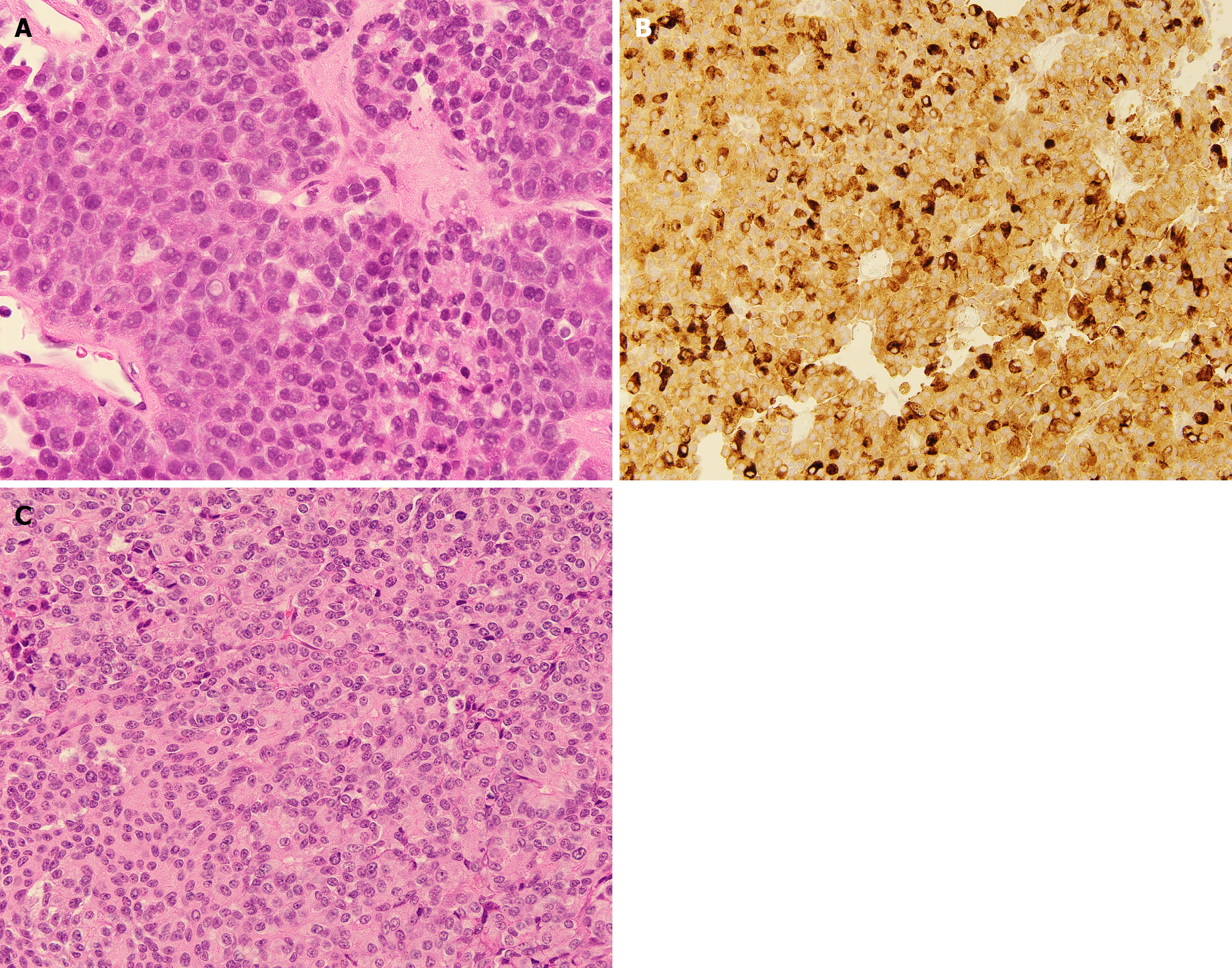
Figure 1 Representative histopathology and immunohistochemical staining of pancreatic acinar cell carcinoma and pancreatoblastoma.
A: Pancreatic acinar cell carcinoma (PACC) patient, hematoxylin and eosin 40 ×: The tumor is composed of sheet-like epithelioid cells, some of them forming acinar structures; B: PACC patient immunohistochemistry 20 ×: The tumor demonstrates cytoplasmic positivity of trypsin. A diagnostic feature of PACC; C: Pancreatoblastoma patient, hematoxylin and eosin 40 ×: Tumor consists of epithelioid cells, some of which form acinar structures with a central lumen.
- Citation: Skorupan N, Sperling D, Nutting C, Miettinen M, Cohn A, Alewine C. Use of yttrium-90 radioembolization to control liver metastases in pancreatic acinar cell carcinoma and pancreatoblastoma: Two case reports. World J Gastroenterol 2026; 32(8): 113861
- URL: https://www.wjgnet.com/1007-9327/full/v32/i8/113861.htm
- DOI: https://dx.doi.org/10.3748/wjg.v32.i8.113861