
















©The Author(s) 2026.
World J Gastroenterol. Feb 28, 2026; 32(8): 113299
Published online Feb 28, 2026. doi: 10.3748/wjg.v32.i8.113299
Published online Feb 28, 2026. doi: 10.3748/wjg.v32.i8.113299
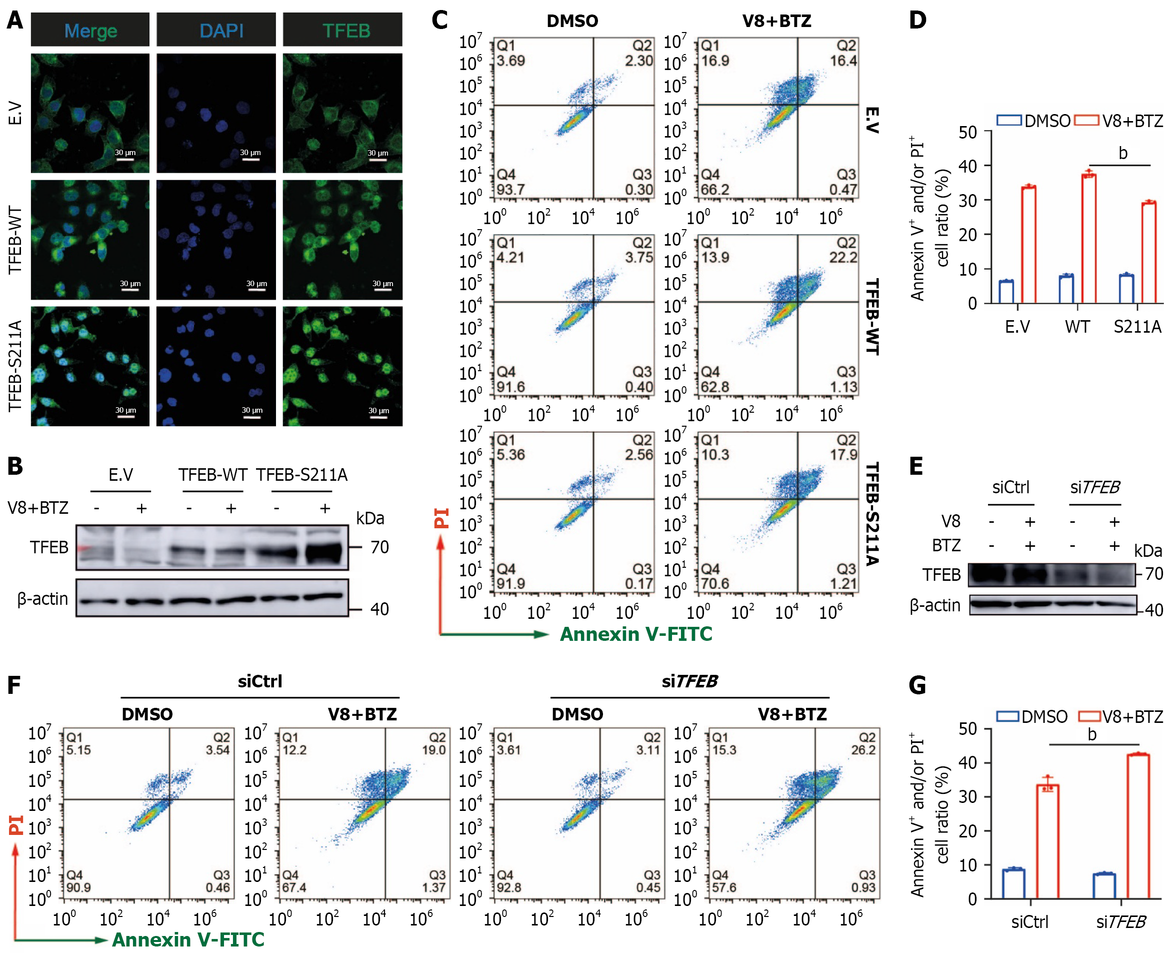
Figure 5 Role of transcription factor EB in cell apoptosis induced by V8 and bortezomib.
A: MGC-803 cells were transiently transfected with empty vector, wild-type, and S211A mutant form of transcription factor EB (TFEB) plasmids. Representative images of immunofluorescence assay are shown for analysis of cellular TFEB localization (scale bar: 30 μm); B-D: MGC-803 cells were transiently transfected with indicated plasmids (B), followed by treatment of V8 (9 μM) together with bortezomib (BTZ) (100 nM) for 24 hours. Representative images (C) and quantitative results (D) are shown; E: TFEB expression was examined by western blotting after transfection of specific small interfering RNA in MGC-803 cells; F and G: MGC-803 cells were treated by V8 (9 μM) combined with BTZ (100 nM) for 24 hours. Representative images (F) and quantified results (G) of cell apoptosis analysis are shown. bP < 0.01. DAPI: 4’,6-Diamidino-2-phenylindole; TFEB: Transcription factor EB; EV: Empty vector; WT: Wild-type; DMSO: Dimethyl sulfoxide; BTZ: Bortezomib; V-FITC: Annexin V-fluorescein isothiocyanate; PI: Propidium iodide; siCtrl: Negative control small interfering RNA; siTFEB: Small interfering RNA targeting TFEB.
- Citation: Li SC, Shao SZ, Zhang YH, Zhou Y, Shang WT, Gao Y, He QB, Guo QL, Guo CY, Zhang XB. Wogonin derivative V8 enhances bortezomib efficacy in gastric carcinoma by disrupting lysosome-mediated drug resistance. World J Gastroenterol 2026; 32(8): 113299
- URL: https://www.wjgnet.com/1007-9327/full/v32/i8/113299.htm
- DOI: https://dx.doi.org/10.3748/wjg.v32.i8.113299