
















Copyright: ©Author(s) 2026.
World J Gastroenterol. Dec 28, 2026; 32(48): 123215
Published online Dec 28, 2026. doi: 10.3748/wjg.123215
Published online Dec 28, 2026. doi: 10.3748/wjg.123215
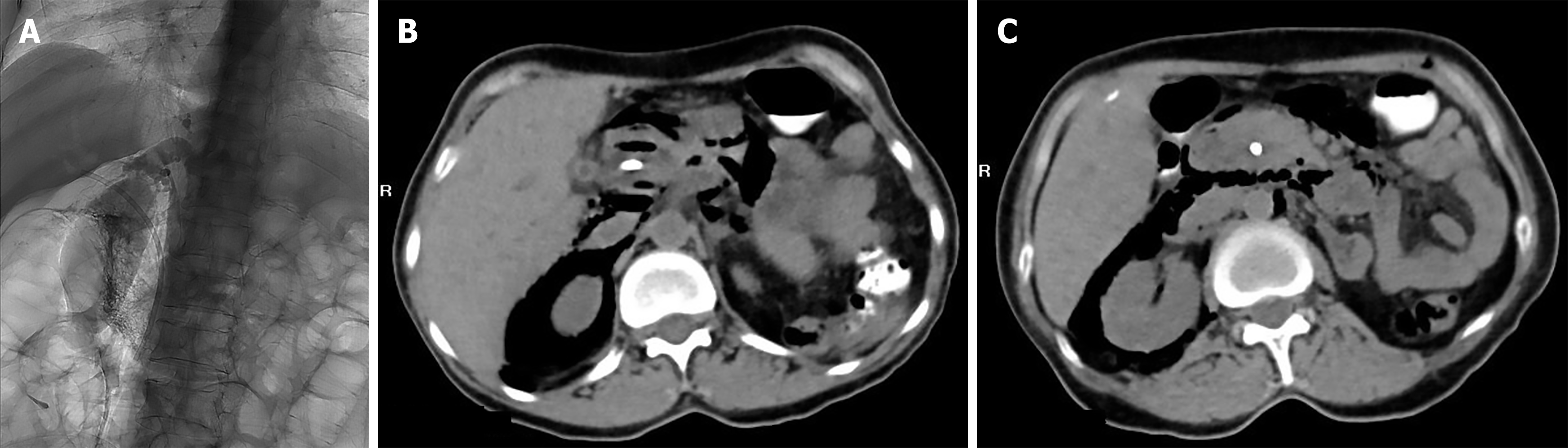
Figure 1 Early suspected retroperitoneal perforation after endoscopic retrograde cholangiopancreatography.
A: Fluoroscopic image during endoscopic retrograde cholangiopancreatography showing dilatation of the intra- and extrahepatic bile ducts, with an approximately 1.2 cm filling defect in the mid-to-upper common bile duct. During gallstone extraction, contrast extravasation was observed and the stone could no longer be clearly delineated; B and C: Axial computed tomography images obtained the following day showing pneumobilia and extensive retroperitoneal extraluminal gas with mediastinal extension, raising suspicion of early retroperitoneal perforation.
- Citation: Danzeng SL, Chen YH, Zha X, Wang Q. Two-stage complications after endoscopic retrograde cholangiopancreatography comprising early retroperitoneal perforation and delayed biliary stent migration: A case report. World J Gastroenterol 2026; 32(48): 123215
- URL: https://www.wjgnet.com/1007-9327/full/v32/i48/123215.htm
- DOI: https://dx.doi.org/10.3748/wjg.123215