
















Copyright: ©Author(s) 2026.
World J Gastroenterol. Dec 28, 2026; 32(48): 122778
Published online Dec 28, 2026. doi: 10.3748/wjg.122778
Published online Dec 28, 2026. doi: 10.3748/wjg.122778
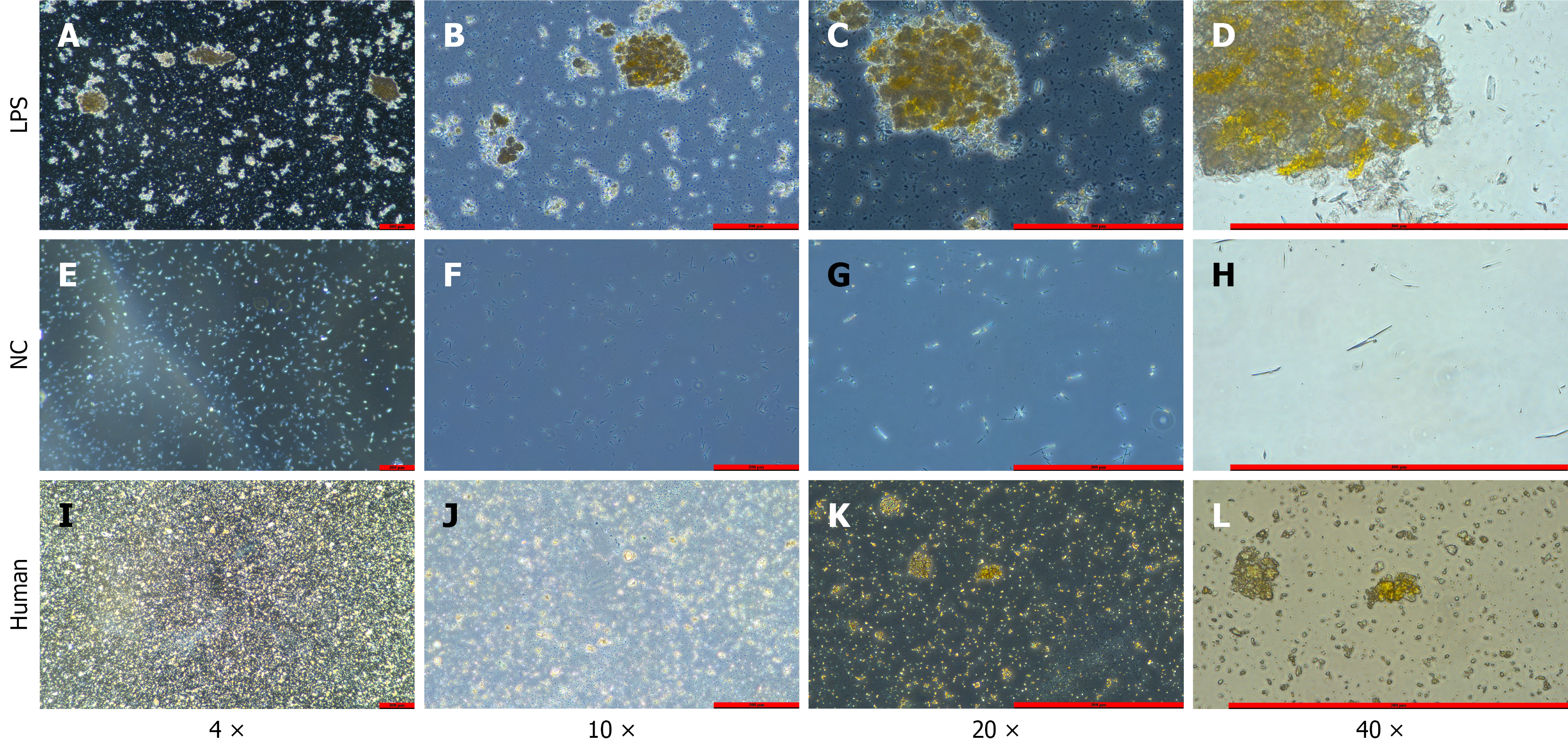
Figure 4 Microscopic bile smears.
A-D: Experimental group (LPS-treated) bile sediment smears at increasing magnifications, showing clearly visible yellow stone-like crystal aggregates with increasing detail at higher magnification; E-H: Control group bile smears after centrifugation at magnifications of 4 ×, 10 ×, 20 ×, and 40 ×, respectively, showing no crystals; I-L: Smears of ground clinical hepatolithiasis stone specimens at the same magnifications, showing yellow crystal morphology essentially consistent with that of the LPS group. LPS: Lipopolysaccharide; NC: Negative control.
- Citation: Shi JL, Muhammad T, He TT, Lin ZY, Zhang TG, Liu SY, Yang XL, Yang Y, Hu L, Ran P, Numan M, Li J. Establishment of a Sprague-Dawley rat model of hepatolithiasis based on common bile duct catheterization. World J Gastroenterol 2026; 32(48): 122778
- URL: https://www.wjgnet.com/1007-9327/full/v32/i48/122778.htm
- DOI: https://dx.doi.org/10.3748/wjg.122778