
















Copyright: ©Author(s) 2026.
World J Gastroenterol. Dec 28, 2026; 32(48): 122778
Published online Dec 28, 2026. doi: 10.3748/wjg.122778
Published online Dec 28, 2026. doi: 10.3748/wjg.122778
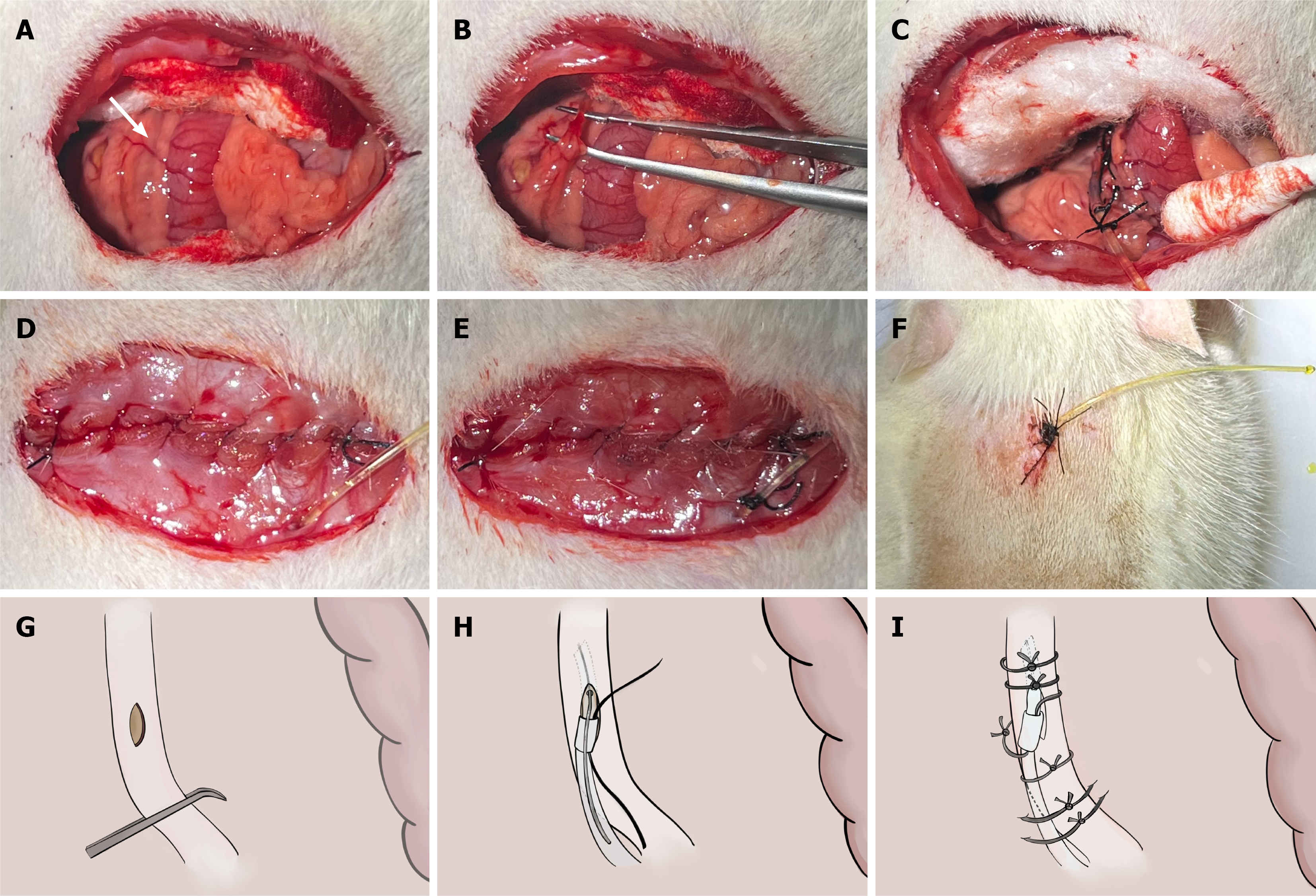
Figure 3 Common bile duct catheterization: Operative steps.
A: Exposure of the common bile duct (white arrow); the duct runs alongside the portal vein and hepatic artery; B: Blunt dissection of the common bile duct using microsurgical forceps; C: Catheter in situ with initial fixation sutures in place and bile flowing freely from the tube; D: Abdominal closure; the catheter passes through the peritoneum and muscle layer; E: Catheter fixed to the abdominal wall before skin closure; F: External catheter externalized and fixed to the back of the neck, leaving 3 cm of the tube exposed; G: Schematic of a 1-mm-long longitudinal incision in the wall of the common bile duct made using a 26-G needle; H: Schematic of insertion of the silicone tube through the incision in the bile duct using a curved clamp while the guidewire is removed; I: Schematic of the final fixation configuration: Bile duct sutures, sleeve suture to the mesentery, and back fixation suture to the mesentery.
- Citation: Shi JL, Muhammad T, He TT, Lin ZY, Zhang TG, Liu SY, Yang XL, Yang Y, Hu L, Ran P, Numan M, Li J. Establishment of a Sprague-Dawley rat model of hepatolithiasis based on common bile duct catheterization. World J Gastroenterol 2026; 32(48): 122778
- URL: https://www.wjgnet.com/1007-9327/full/v32/i48/122778.htm
- DOI: https://dx.doi.org/10.3748/wjg.122778