
















Copyright: ©Author(s) 2026.
World J Gastroenterol. Dec 21, 2026; 32(47): 122949
Published online Dec 21, 2026. doi: 10.3748/wjg.122949
Published online Dec 21, 2026. doi: 10.3748/wjg.122949
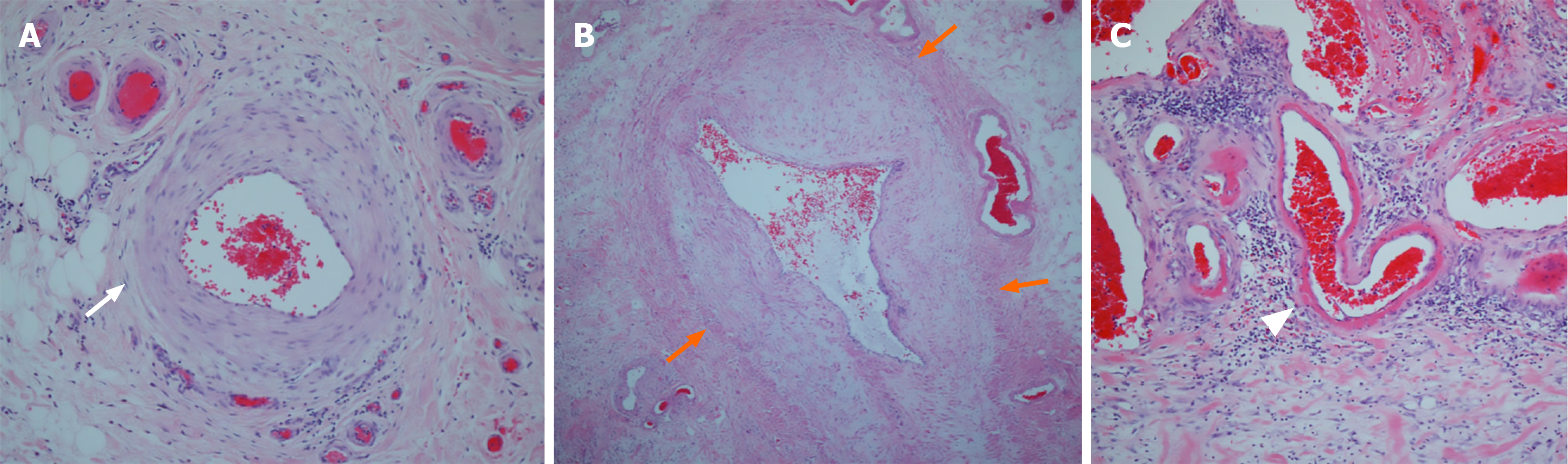
Figure 5 Histopathologic findings of the resected colon consistent with idiopathic myointimal hyperplasia of mesenteric veins.
A: A mesenteric artery showing a preserved normal wall architecture without intimal thickening (white arrow), × 100; B: A mesenteric vein demonstrating marked myointimal hyperplasia with near-complete luminal obliteration (orange arrows), × 100; C: Small venules exhibiting subendothelial fibrin deposition, seen as eosinophilic material beneath the endothelium (arrowhead), × 40. These findings are characteristic of idiopathic myointimal hyperplasia of mesenteric veins, highlighting selective venous involvement with arterial sparing.
- Citation: Lee JS, Kim EY, Kwon JG, Bae JH, Kim M, Yang CS, Yu SH, Cho CH, Jo HH. Idiopathic myointimal hyperplasia of mesenteric veins mimicking steroid-refractory ulcerative colitis: A case report. World J Gastroenterol 2026; 32(47): 122949
- URL: https://www.wjgnet.com/1007-9327/full/v32/i47/122949.htm
- DOI: https://dx.doi.org/10.3748/wjg.122949