
















Copyright: ©Author(s) 2026.
World J Gastroenterol. Dec 21, 2026; 32(47): 122949
Published online Dec 21, 2026. doi: 10.3748/wjg.122949
Published online Dec 21, 2026. doi: 10.3748/wjg.122949
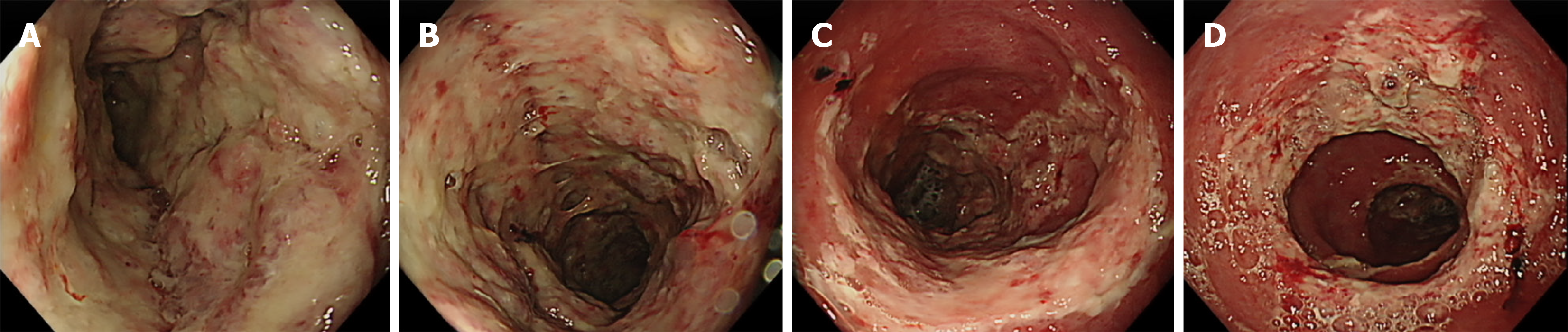
Figure 3 Follow-up sigmoidoscopy demonstrating marked progression of ulcerative lesions.
Follow-up sigmoidoscopy performed for hematochezia showed marked worsening of colonic inflammation despite ongoing medical therapy. The transverse colon and descending colon demonstrated extensive mucosal ulceration with irregular, deep ulcers covered by thick exudate. Similar findings were observed in the sigmoid colon and rectum, where ulcerations appeared more extensive and deeper, with areas of mucosal denudation and suspected vascular exposure. These findings indicate severe progression of disease activity despite corticosteroid and subsequent filgotinib therapy. A: Transverse colon; B: Descending colon; C: Sigmoid colon; D: Rectum.
- Citation: Lee JS, Kim EY, Kwon JG, Bae JH, Kim M, Yang CS, Yu SH, Cho CH, Jo HH. Idiopathic myointimal hyperplasia of mesenteric veins mimicking steroid-refractory ulcerative colitis: A case report. World J Gastroenterol 2026; 32(47): 122949
- URL: https://www.wjgnet.com/1007-9327/full/v32/i47/122949.htm
- DOI: https://dx.doi.org/10.3748/wjg.122949