
















Copyright: ©Author(s) 2026.
World J Gastroenterol. Dec 21, 2026; 32(47): 120094
Published online Dec 21, 2026. doi: 10.3748/wjg.v32.i47.120094
Published online Dec 21, 2026. doi: 10.3748/wjg.v32.i47.120094
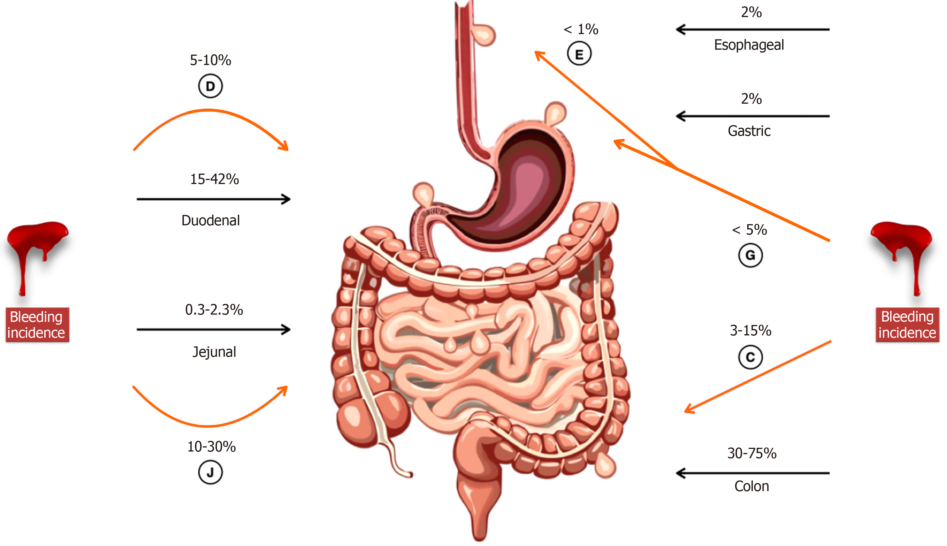
Figure 1 Anatomic distribution of diverticula and site-specific bleeding incidence.
Black arrows show gastrointestinal (GI) diverticula incidence. Incidence of diverticula bleeding is highlighted with a orange arrow. Jejunal diverticula accounts for a disproportionately higher bleeding rate (10%-30%). This distribution highlights the clinical relevance of jejunal diverticular disease in the assessment of unspecific GI bleeding. D: Duodenal; J: Jejunal; E: Esophageal; G: Gastric; C: Colon.
- Citation: Anaya-Prado R, Dominguez-Soto AV, Osorio-Medina KN, Ortiz-Vera RI, Gonzalez-Martinez D, Anaya-Fernández R, Portillo-Mendez R, Anaya-Fernández MM, Azcona-Ramirez CC, Guerrero-Palomera MA, Peña-Cuellar AJ, Soto-Hintze J, Reyna-Rodriguez B. Jejunal diverticula bleeding: A challenging diagnosis entity. World J Gastroenterol 2026; 32(47): 120094
- URL: https://www.wjgnet.com/1007-9327/full/v32/i47/120094.htm
- DOI: https://dx.doi.org/10.3748/wjg.v32.i47.120094