
















Copyright: ©Author(s) 2026.
World J Gastroenterol. Dec 14, 2026; 32(46): 121572
Published online Dec 14, 2026. doi: 10.3748/wjg.121572
Published online Dec 14, 2026. doi: 10.3748/wjg.121572
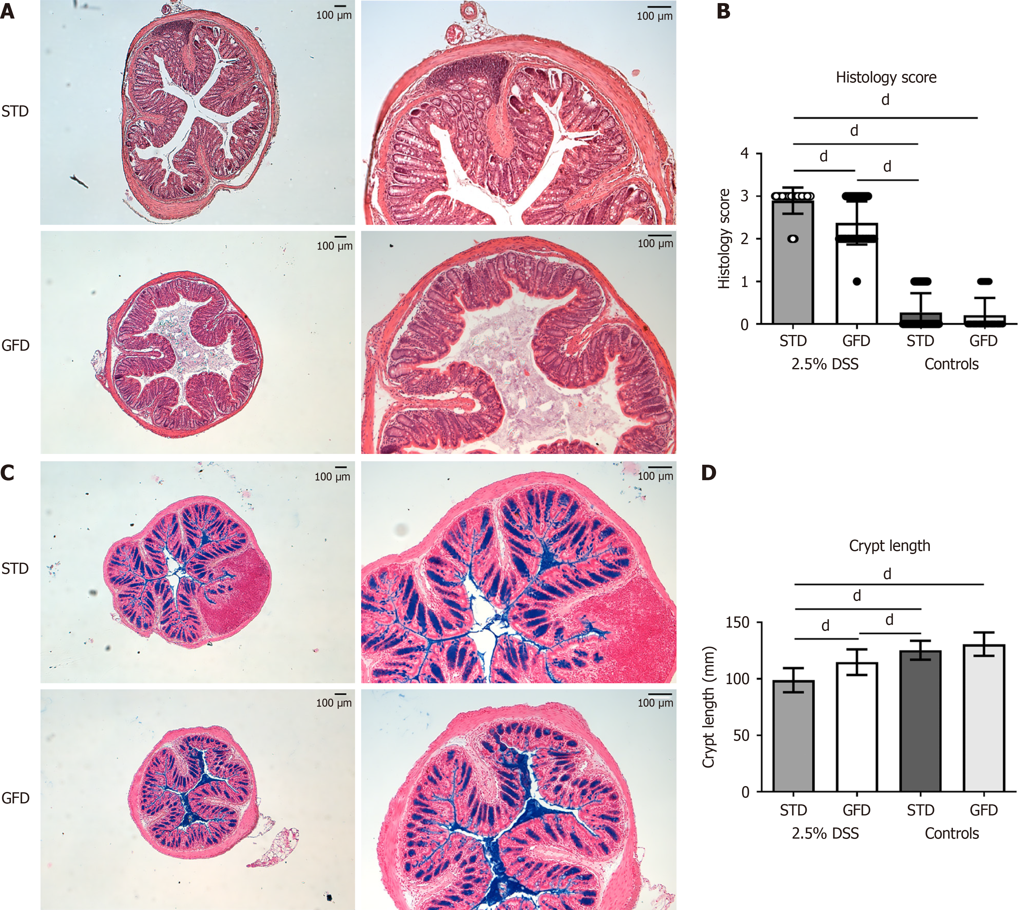
Figure 4 Colon morphology and histology of the dextran sulfate sodium-treated BALB/c mice fed gluten-free diet or standard diet.
A: Representative hematoxylin and eosin-stained colon sections from BALB/c mice treated with 2.5% dextran sulfate sodium (DSS) for 7 days and fed the gluten-free diet (GFD) or standard diet (STD) (n = 10), magnification 5 × or 10 ×; B: Histology score evaluating colonic mucosa in GFD- and STD-fed BALB/c mice (P < 0.0001, n = 10) after 7 days of 2.5% DSS exposure, including untreated controls; C: Representative histological specimens documenting mucin in colon and goblet cells within colonic epithelium (stained by Alcian blue and nuclear fast red) of BALB/c mice treated for 7 days with 2.5% DSS and fed GFD or STD (n = 10), magnification 5 × or 10 ×; D: Statistically significant (P < 0.0001) preservation of crypt length in GFD vs STD BALB/c mice (n = 10) exposed to 2.5% DSS. Example of two independent experiments. Data are expressed as mean ± SD. dP < 0.0001. DSS: Dextran sulfate sodium; STD: Standard diet; GFD: Gluten-free diet.
- Citation: Funda D, Drabonova B, Golias J, Tejklova T, Cisarova R, Bartosova J, Drastich P. Gluten-free diet reduces severity of dextran sulfate sodium-induced colitis in BALB/c mice. World J Gastroenterol 2026; 32(46): 121572
- URL: https://www.wjgnet.com/1007-9327/full/v32/i46/121572.htm
- DOI: https://dx.doi.org/10.3748/wjg.121572