
















Copyright: ©Author(s) 2026.
World J Gastroenterol. Dec 7, 2026; 32(45): 121242
Published online Dec 7, 2026. doi: 10.3748/wjg.121242
Published online Dec 7, 2026. doi: 10.3748/wjg.121242
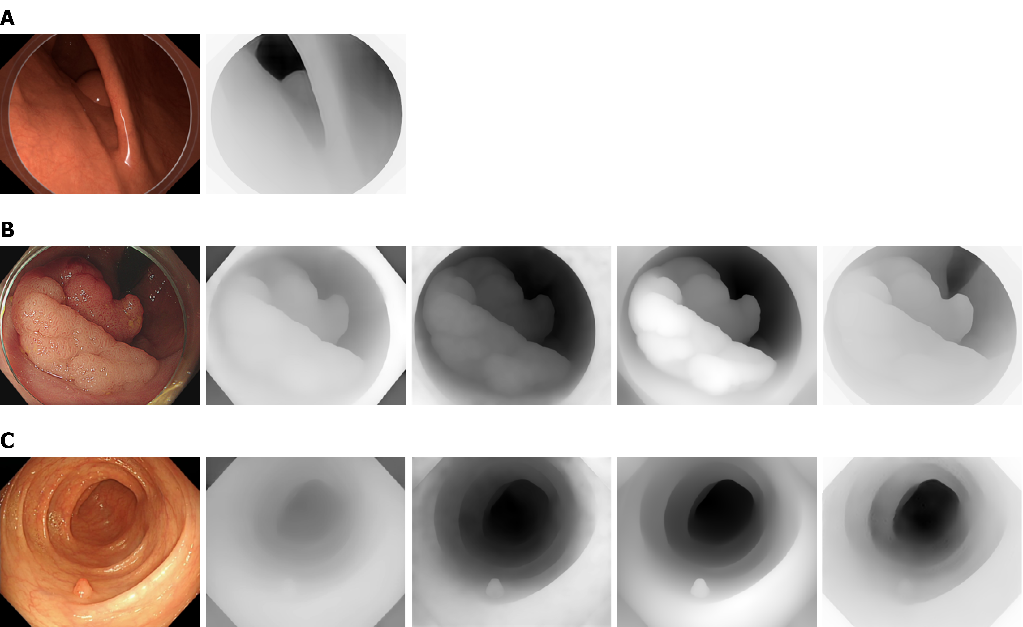
Figure 2 Visual inspection indicates that all models generate plausible representations of the depth structure.
A: The first row shows an image from the virtual colonoscopy alongside the predicted depth map of the fine-tuned Depth Anything V2 model. Depth is encoded in grayscale, with black indicating greater distance and white indicating closer proximity to the camera; B: The second row presents a representative whitelight endoscopic image from the endoscopic submucosal dissection cohort, accompanied by depth map predictions generated by Apple Depth Pro, UniK3D, Depth Anything V2 (nonfinetuned), as well as Depth Anything V2 (fine-tuned). Qualitatively, all models provide a reasonable representation of the depth structure in the image; C: The third row illustrates a representative whitelight endoscopic image from the external dataset, together with the corresponding depth map predictions from Apple Depth Pro, UniK3D, Depth Anything V2 (non-fine-tuned), and Depth Anything V2 (fine-tuned).
- Citation: Schulz D, Ebigbo A, Holley E, Nagl S, Nichiporyk O, Probst A, Scheppach MW, Römmele C, Roser D, Zellmer S, Messmann H. Domain specific training with three-dimensional virtual colonoscopy images improves artificial intelligence based automated size prediction for large lesions. World J Gastroenterol 2026; 32(45): 121242
- URL: https://www.wjgnet.com/1007-9327/full/v32/i45/121242.htm
- DOI: https://dx.doi.org/10.3748/wjg.121242