
















Copyright: ©Author(s) 2026.
World J Gastroenterol. Nov 28, 2026; 32(44): 121710
Published online Nov 28, 2026. doi: 10.3748/wjg.121710
Published online Nov 28, 2026. doi: 10.3748/wjg.121710
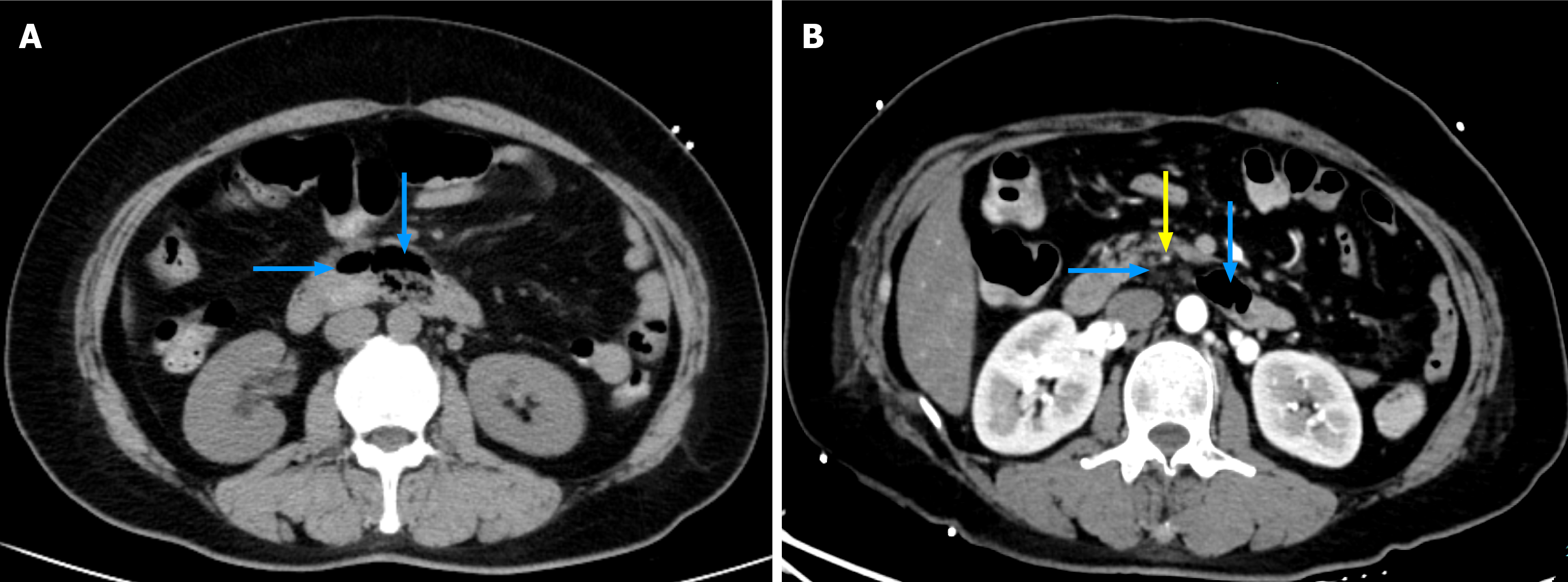
Figure 1 Enhanced abdominal computed tomography imaging.
A: Non-contrast computed tomography scan showing multiple duodenal diverticula; B: Arterial phase contrast imaging revealing a suspected arterial vessel coursing through the wall of one diverticulum. Blue arrow: Duodenal diverticula in the horizontal segment of the duodenum; yellow arrow: Suspected intradiverticular arterial vessel.
- Citation: Shen YJ, Song JJ, Liu XQ, Hu FL. Phenylephrine-provoked endoscopy reveals an occult Dieulafoy’s lesion in a duodenal diverticulum: A case report and review of literature. World J Gastroenterol 2026; 32(44): 121710
- URL: https://www.wjgnet.com/1007-9327/full/v32/i44/121710.htm
- DOI: https://dx.doi.org/10.3748/wjg.121710