
















Copyright: ©Author(s) 2026.
World J Gastroenterol. Nov 14, 2026; 32(42): 121690
Published online Nov 14, 2026. doi: 10.3748/wjg.121690
Published online Nov 14, 2026. doi: 10.3748/wjg.121690
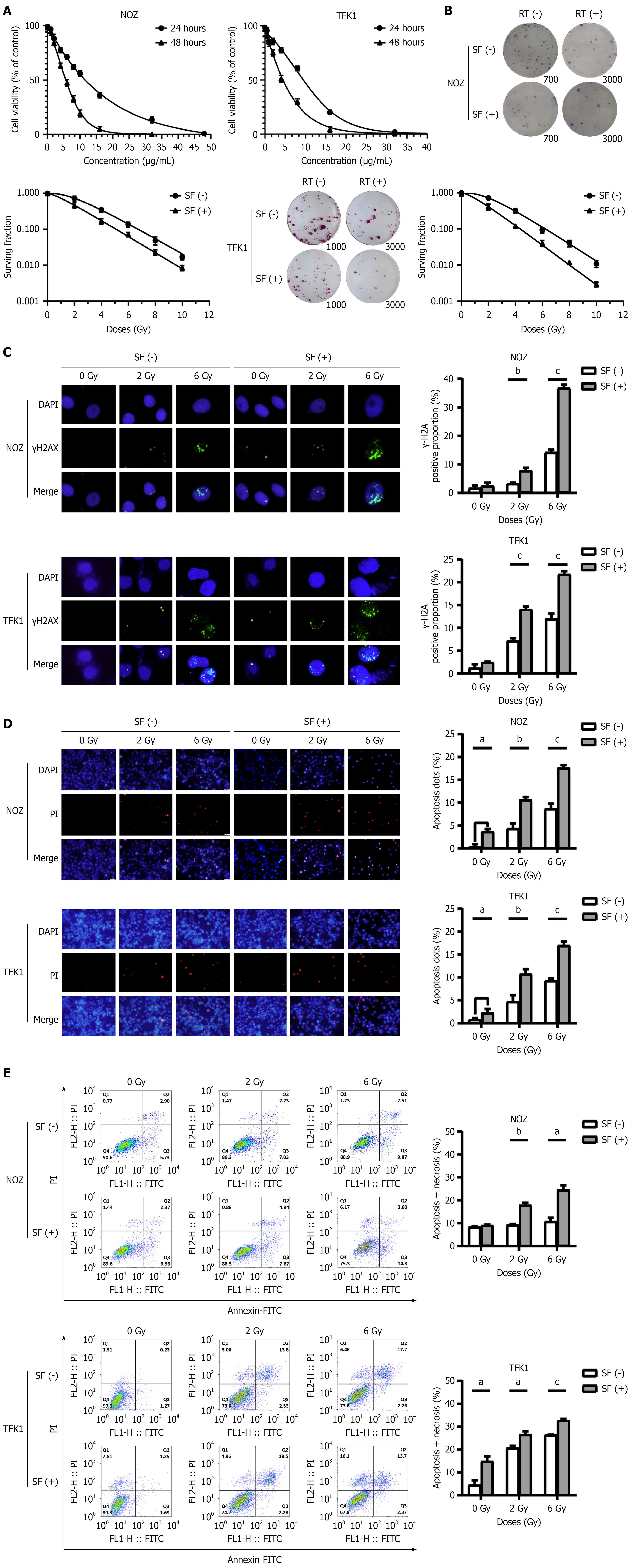
Figure 1 Surufatinib is associated with enhanced radiosensitivity of NOZ and TFK-1 and increased DNA damage.
A: Cell viability assays were performed by Cell Counting Kit-8. Concentrations are shown in μg/mL. The anti-proliferative effect of surufatinib on NOZ and TFK-1 cells; B: Representative colony formation staining images and survival curves of NOZ and TFK-1 with different treatments. The survival curves were plotted using the multi-target single-hit model; C: Surufatinib increased the fluorescence foci of γ-H2AX in tumor cells after radiotherapy. Representative fluorescence foci images and quantitative detection of γ-H2AX foci in NOZ and TFK-1 cells following different treatments; scoring was performed blindly by two investigators; D and E: Apoptosis assessment by flow cytometry in NOZ and TFK-1 cells following different treatments. Apoptotic rates were calculated as the percentages of annexin-V FITC-positive cells. All columns indicate mean and error bars indicate SEM from three independent experiments. aP < 0.05, bP < 0.01, cP < 0.001. SF: Surufatinib; RT: Radiotherapy.
- Citation: Huang A, Cao Y, Li B, Xiao J, Zhan SD, Wang NY, Mei XP, Zhao SZ, Han J, Xiao Y, Wang LF, Ma H. Surufatinib achieves radiosensitivity in cholangiocarcinoma by suppressing GINS4 expression to induce Ca2+/cAMP signaling activation. World J Gastroenterol 2026; 32(42): 121690
- URL: https://www.wjgnet.com/1007-9327/full/v32/i42/121690.htm
- DOI: https://dx.doi.org/10.3748/wjg.121690