
















Copyright: ©Author(s) 2026.
World J Gastroenterol. Nov 14, 2026; 32(42): 120663
Published online Nov 14, 2026. doi: 10.3748/wjg.120663
Published online Nov 14, 2026. doi: 10.3748/wjg.120663
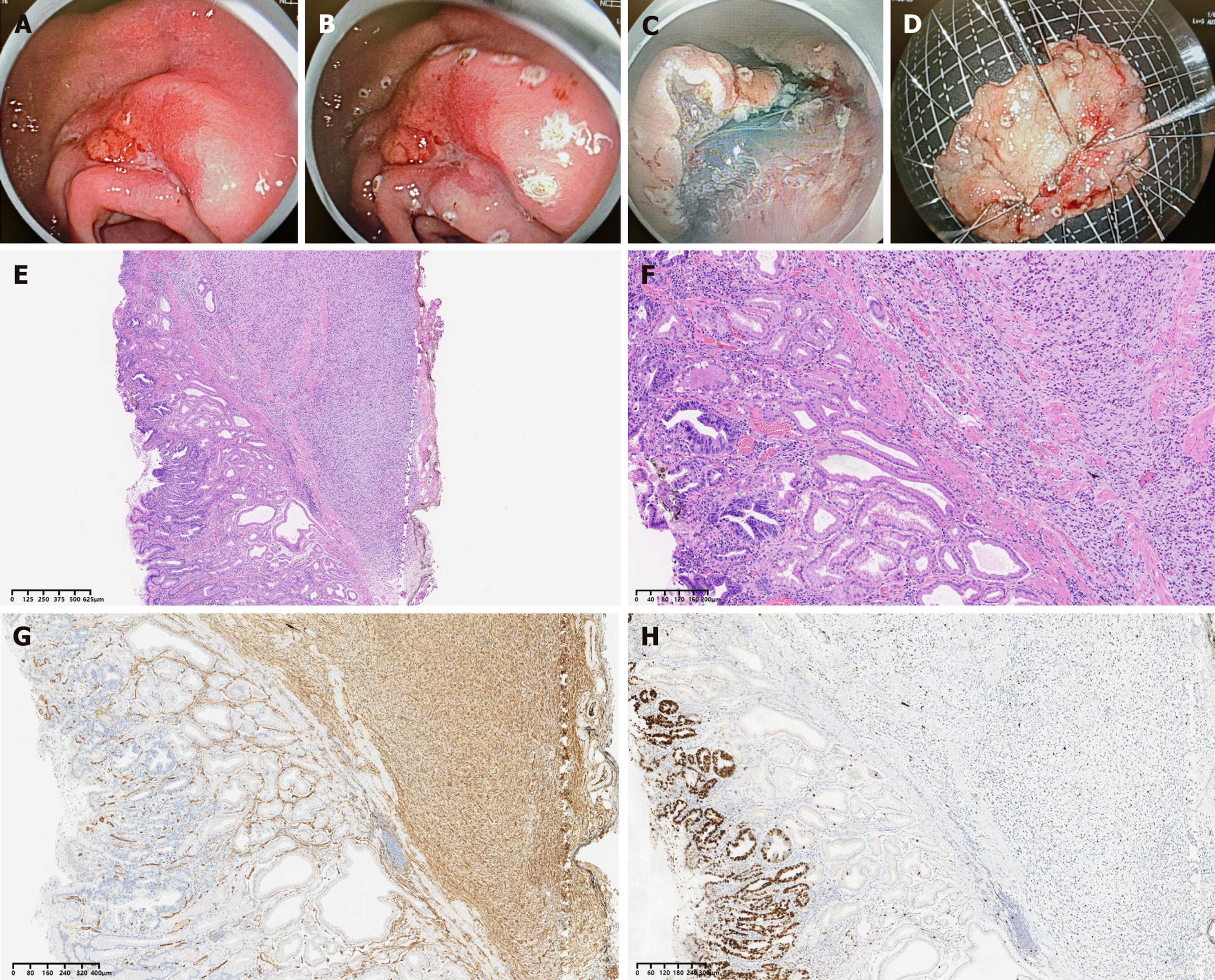
Figure 3 Endoscopic and pathological findings of an inflammatory fibroid polyp concurrent with early gastric cancer.
A: White light endoscopy showing a gastric antrum lesion with erythema and an irregular, rough mucosal surface; B and C: Endoscopic resection of the lesion via endoscopic submucosal dissection; D: The resected specimen measuring 50 mm × 35 mm; E: Low-power view (hematoxylin and eosin staining, × 40) of inflammatory fibroid polyp (IFP) overlying adenocarcinoma tissue; F: High-power view (hematoxylin and eosin staining, × 200) of IFP adjacent to early gastric cancer foci; G: CD34 immunohistochemical staining (× 200) showing diffuse expression in spindle tumor cells of IFP; H: Ki67 immunohistochemical staining (× 200) with a high proliferation index in the adenocarcinoma region and a low index in the IFP region.
- Citation: Wang CH, Liu F, Ji XH, Li LS, Chai NL. Clinical characteristics and endoscopic management of inflammatory fibroid polyp of the gastrointestinal tract. World J Gastroenterol 2026; 32(42): 120663
- URL: https://www.wjgnet.com/1007-9327/full/v32/i42/120663.htm
- DOI: https://dx.doi.org/10.3748/wjg.120663