
















Copyright: ©Author(s) 2026.
World J Gastroenterol. Nov 14, 2026; 32(42): 120663
Published online Nov 14, 2026. doi: 10.3748/wjg.120663
Published online Nov 14, 2026. doi: 10.3748/wjg.120663
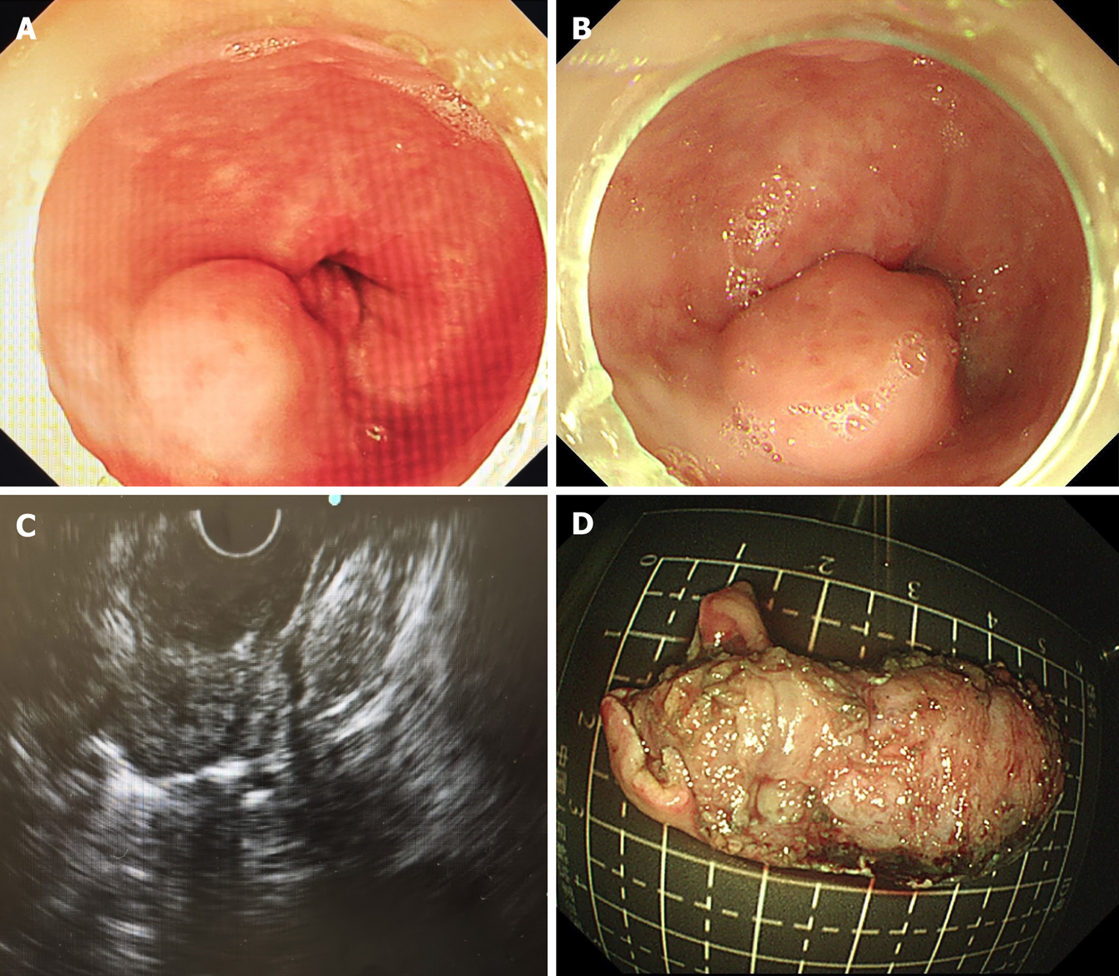
Figure 1 Endoscopic and endoscopic ultrasound features of an esophageal inflammatory fibroid polyp.
A: A large tumor in the esophagus; B: Close observation revealed that this lesion was located above the dentate line; C: A hyperechoic tumor with slightly heterogeneous internal echoes located at the esophagus; D: The resected specimen was 50 mm × 30 mm.
- Citation: Wang CH, Liu F, Ji XH, Li LS, Chai NL. Clinical characteristics and endoscopic management of inflammatory fibroid polyp of the gastrointestinal tract. World J Gastroenterol 2026; 32(42): 120663
- URL: https://www.wjgnet.com/1007-9327/full/v32/i42/120663.htm
- DOI: https://dx.doi.org/10.3748/wjg.120663