
















Copyright: ©Author(s) 2026.
World J Gastroenterol. Nov 14, 2026; 32(42): 120411
Published online Nov 14, 2026. doi: 10.3748/wjg.120411
Published online Nov 14, 2026. doi: 10.3748/wjg.120411
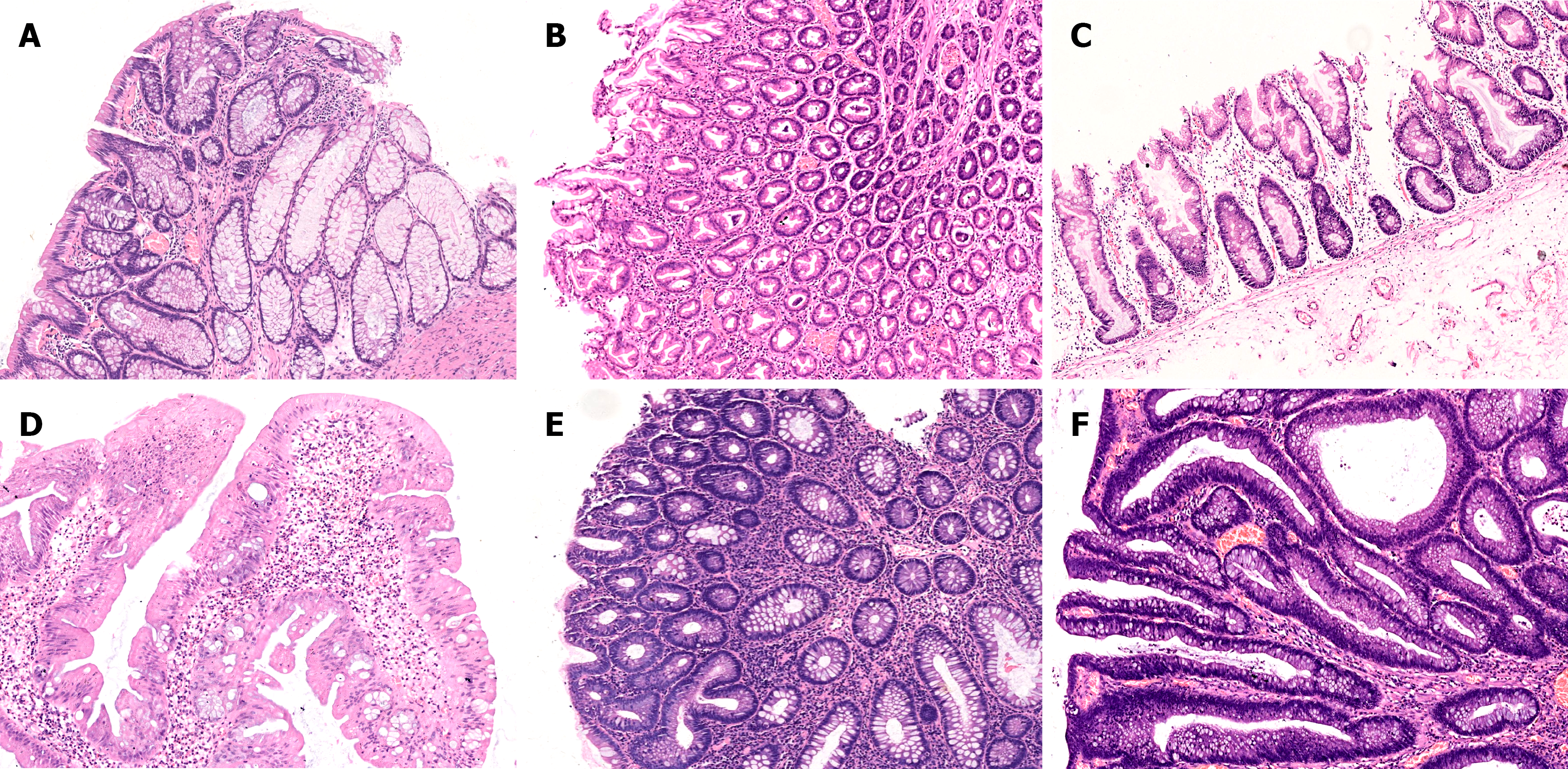
Figure 1 Pathomorphological features of different types of colorectal polyp lesions.
A: Similar to normal crypt morphology, but primarily composed of goblet cells with small, uniform nuclei located at the base (goblet cell-rich hyperplastic polyp, hematoxylin-eosin, original magnification 100 ×); B: The surface of the crypts exhibits serrated architectures, with the cross-section displaying a star-like pattern, and small droplet-like mucus can be observed scattered within the epithelial cells (microvesicular hyperplastic polyp, hematoxylin-eosin, original magnification 40 ×); C: The serrated crypts are prominently displayed, extending deeply into the crypts with dilation and branching, giving the gland a distinctive ‘L’-shaped configuration (sessile serrated lesion, hematoxylin-eosin, original magnification 100 ×); D: Serrated or fissure-like crypts are observed, with the presence of ectopic crypts. The nuclei exhibit elongation resembling a pencil, and the cytoplasm displays significant eosinophilia (traditional serrated adenoma, hematoxylin-eosin, original magnification 100 ×); E: The glandular tissue exhibits an irregular tubular pattern with a crowded arrangement of cells and deeply stained nuclei (tubular adenoma, hematoxylin-eosin, original magnification 100 ×); F: The glandular tissue exhibits an irregular tubular and villous configuration, with cells demonstrating atypia and deeply stained nuclei (tubulovillous adenoma, hematoxylin-eosin, original magnification 100 ×).
- Citation: Hu H, Zhao SB, Zhao YD, Sui XY, Zhang S, Wei JH, Liu XF, Shi YC, Tang L, Zhang BB, Feng Z, Cheng W, Zhu MH, Zhu Z, Li ZS, He MX, Bai Y, Zhang J. Low accuracy and interobserver/intraobserver agreement for colorectal sessile serrated lesions: A multicenter diagnostic study. World J Gastroenterol 2026; 32(42): 120411
- URL: https://www.wjgnet.com/1007-9327/full/v32/i42/120411.htm
- DOI: https://dx.doi.org/10.3748/wjg.120411