
















Copyright: ©Author(s) 2026.
World J Gastroenterol. Nov 7, 2026; 32(41): 121668
Published online Nov 7, 2026. doi: 10.3748/wjg.121668
Published online Nov 7, 2026. doi: 10.3748/wjg.121668
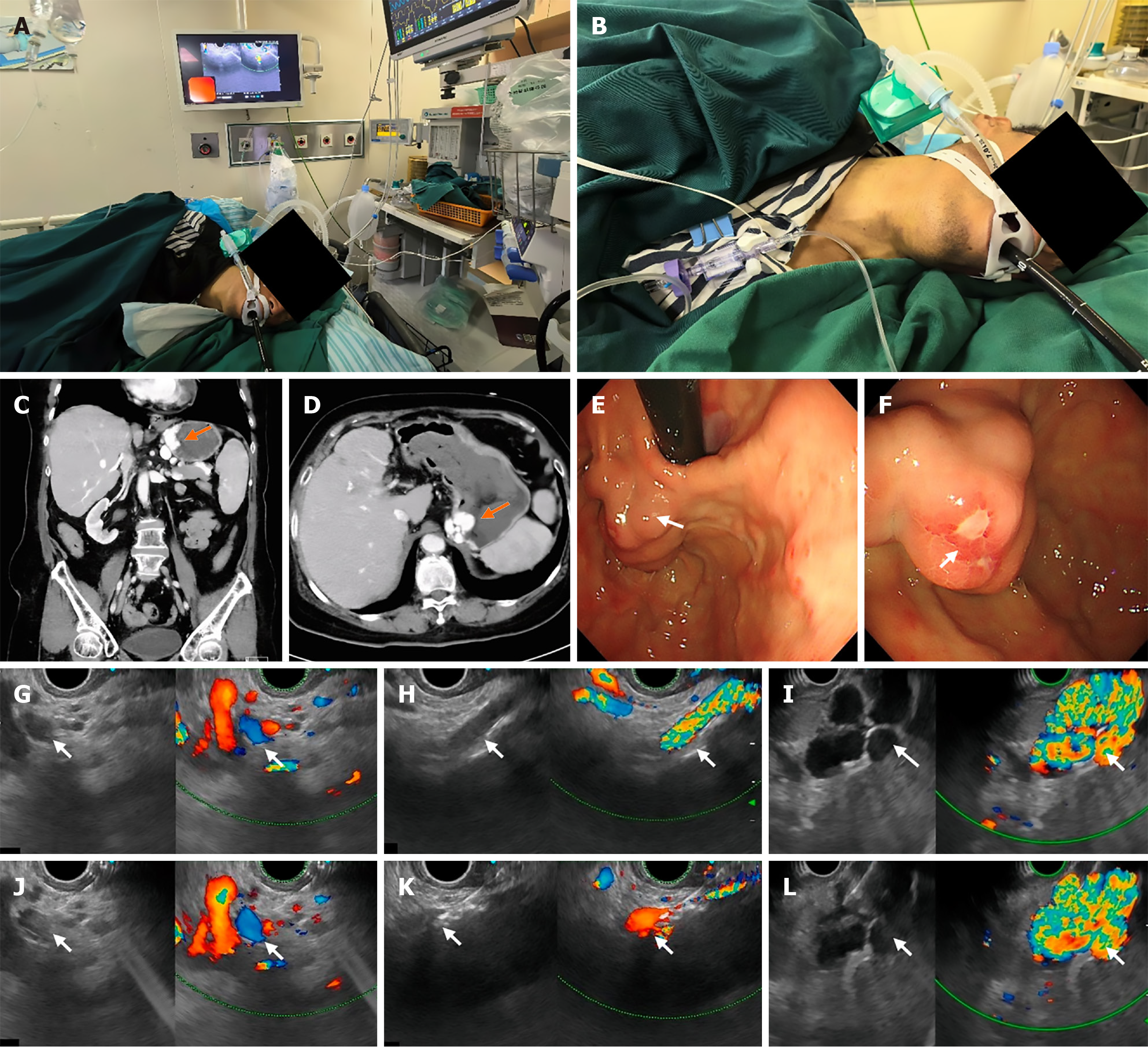
Figure 1 Endoscopic ultrasound-guided portal pressure measurement process.
A: Endoscopic ultrasound (EUS)-guided portal pressure measurement process; B: Zero calibration of the pressure transducer at the right atrial level; C: Gastric fundal varix on coronal computed tomography (CT) imaging; D: Gastric fundal varix on sagittal CT imaging; E: Endoscopic views of gastric fundal varices; F: Endoscopic views of gastric fundal varices; G: Visualization of the main portal vein under EUS, the arrow indicates the main portal vein; H: Visualization of the inferior vena cava under EUS, the arrow indicates the inferior vena cava; I: Visualization of gastric fundal varices under EUS, the arrow indicates gastric fundal varices; J: EUS-guided needle puncture of the main portal vein, the arrow indicates the main portal vein; K: EUS-guided needle puncture of the inferior vena cava, the arrow indicates the inferior vena cava; L: EUS-guided needle puncture of the gastric varix, the arrow indicates gastric fundal varices.
- Citation: Wei W, Xue M, Xu LY. Gastric variceal pressure gradient in patients with large spontaneous shunts: A novel approach to local hemodynamic assessment. World J Gastroenterol 2026; 32(41): 121668
- URL: https://www.wjgnet.com/1007-9327/full/v32/i41/121668.htm
- DOI: https://dx.doi.org/10.3748/wjg.121668