
















Copyright: ©Author(s) 2026.
World J Gastroenterol. Oct 28, 2026; 32(40): 121857
Published online Oct 28, 2026. doi: 10.3748/wjg.121857
Published online Oct 28, 2026. doi: 10.3748/wjg.121857
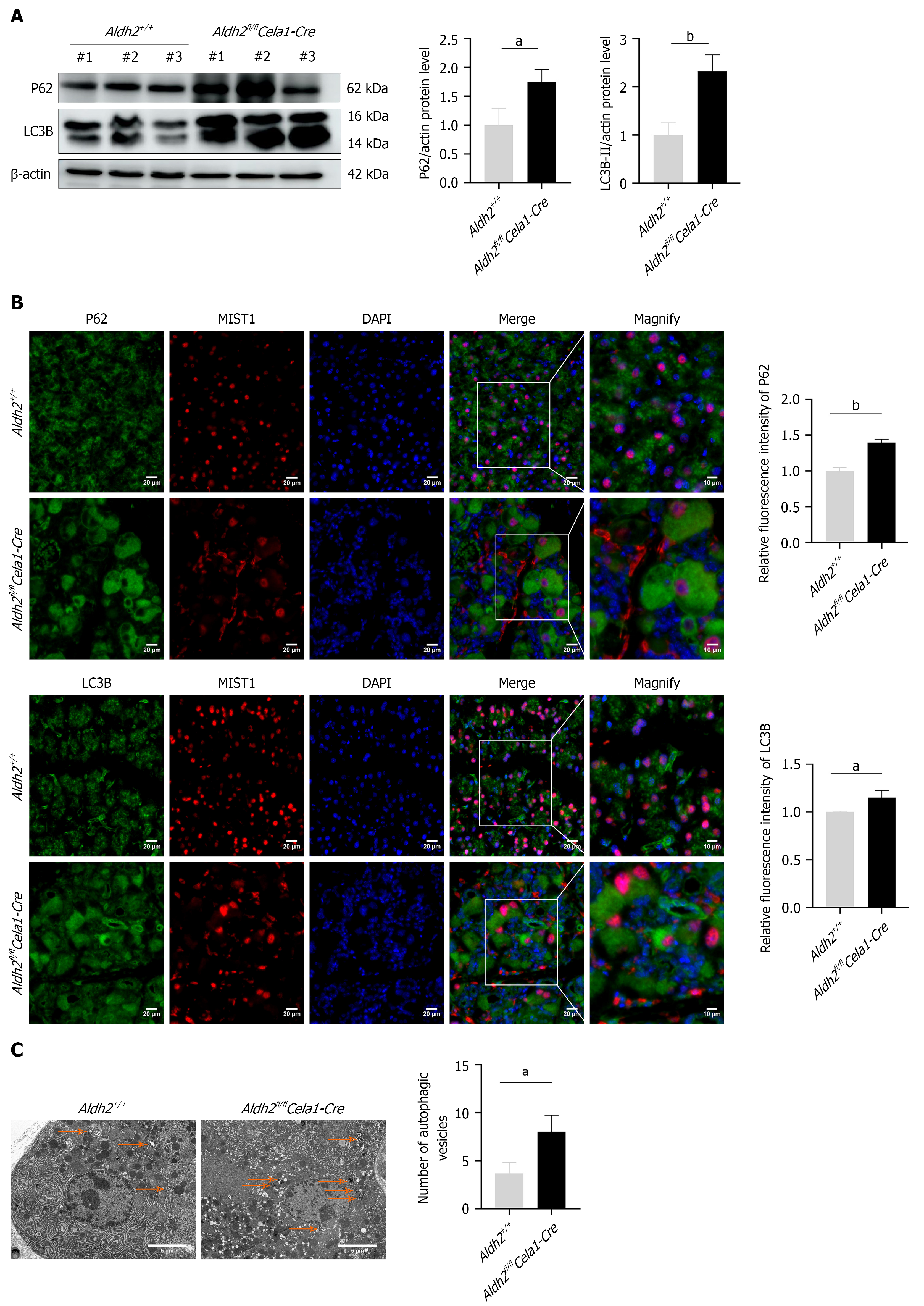
Figure 4 Aldehyde dehydrogenase 2 deficiency in pancreatic acinar cells exacerbated accumulation of autophagic substrates in murine acute pancreatitis models.
Acute pancreatitis (AP) was induced in pancreatic acinar cell-specific aldehyde dehydrogenase 2 knockout mice (Aldh2fl/flCela1-Cre) and the wild-type mice (Aldh2+/+) by seven intraperitoneal injections of caerulein (50 µg/kg). A: The expression of P62 and microtubule-associated protein light chain 3B (LC3B) in pancreatic tissues of Aldh2fl/flCela1-Cre mice and Aldh2+/+ mice by immunoblotting (n = 3 per group); B: Immunofluorescence analysis of P62 and LC3B in acinar cells of pancreatic tissues from Aldh2fl/flCela1-Cre mice and Aldh2+/+ mice with AP (n = 3 per group). Scale bar: 20 μm, magnification, 10 μm. MIST-1 is used as a marker of acinar cells; C: Representative transmission electron microscopy images and quantitative analysis in pancreatic tissue from Aldh2fl/flCela1-Cre mice and Aldh2+/+ mice with AP. Arrows denote autophagic vesicles. Scale bar: 5 μm. aP < 0.05, bP < 0.01. Aldh2fl/flCela1-Cre: Aldehyde dehydrogenase 2 knockout mice.
- Citation: Wu QZ, Shao BZ, Sun HY, Guan KY, Wang Y, Liu ZY, Zhang WG, Linghu EQ. ALDH2 deficiency in pancreatic acinar cells aggravates acute pancreatitis by promoting NLRP3 inflammasome activation through autophagy. World J Gastroenterol 2026; 32(40): 121857
- URL: https://www.wjgnet.com/1007-9327/full/v32/i40/121857.htm
- DOI: https://dx.doi.org/10.3748/wjg.121857