
















Copyright: ©Author(s) 2026.
World J Gastroenterol. Oct 21, 2026; 32(39): 121007
Published online Oct 21, 2026. doi: 10.3748/wjg.121007
Published online Oct 21, 2026. doi: 10.3748/wjg.121007
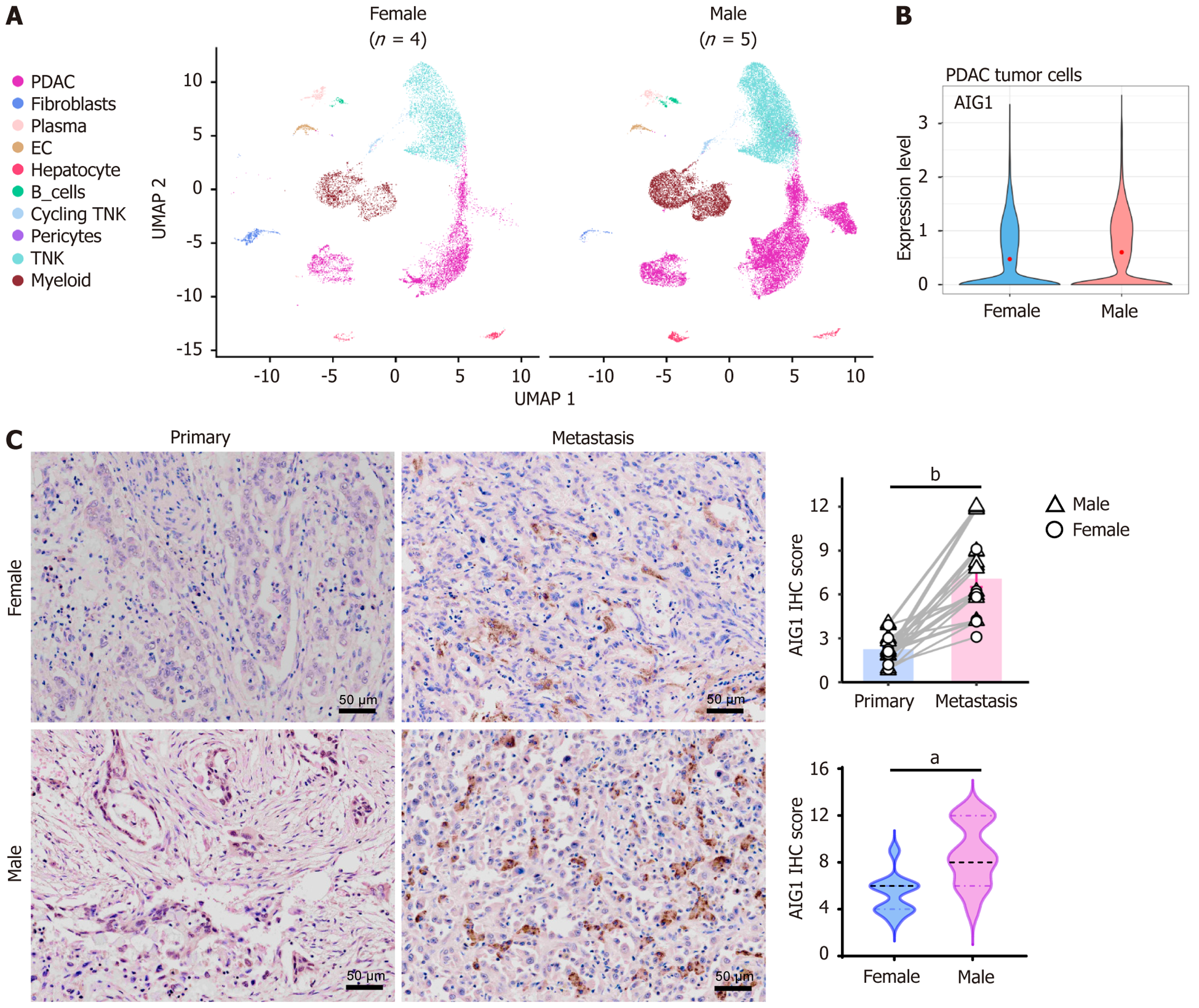
Figure 2 Sex-associated expression pattern of androgen-induced gene 1 in human pancreatic ductal adenocarcinoma liver metastases.
A: Uniform manifold approximation and projection visualization of liver metastatic samples stratified by sex (male, n = 5; female, n = 4) from the public single-cell RNA sequencing dataset; B: Violin plot showing androgen-induced gene 1 (AIG1) expression levels in tumor cells derived from male and female liver metastases. Compared using Wilcoxon rank-sum test, with adjusted P < 0.0001; C: Representative immunohistochemical staining of AIG1 in paired primary pancreatic tumors and matched liver metastatic lesions from patients (n = 24; 13 males and 11 females). Semi-quantification of AIG1 immunohistochemical scores is shown on the right. Comparisons between primary and metastatic tumors were performed using Wilcoxon signed-rank test, and comparisons between male and female metastatic samples were performed using Mann-Whitney U test. aP < 0.01; bP < 0.0001. Scale bar = 50 μm. PDAC: Pancreatic ductal adenocarcinoma; EC: Endothelial cells; TNK: T and natural killer; NK: Natural killer; AIG1: Androgen-induced gene 1; IHC: Immunohistochemical.
- Citation: Luo YZ, Zhao Q, Wang FF, Li JH, Lyu SC, Liu Z, Lang R, He Q. Androgen-induced gene 1 correlates with extracellular vesicle-associated cholesterol metabolism and hepatic metastasis of pancreatic cancer. World J Gastroenterol 2026; 32(39): 121007
- URL: https://www.wjgnet.com/1007-9327/full/v32/i39/121007.htm
- DOI: https://dx.doi.org/10.3748/wjg.121007