
















Copyright: ©Author(s) 2026.
World J Gastroenterol. Oct 21, 2026; 32(39): 120546
Published online Oct 21, 2026. doi: 10.3748/wjg.120546
Published online Oct 21, 2026. doi: 10.3748/wjg.120546
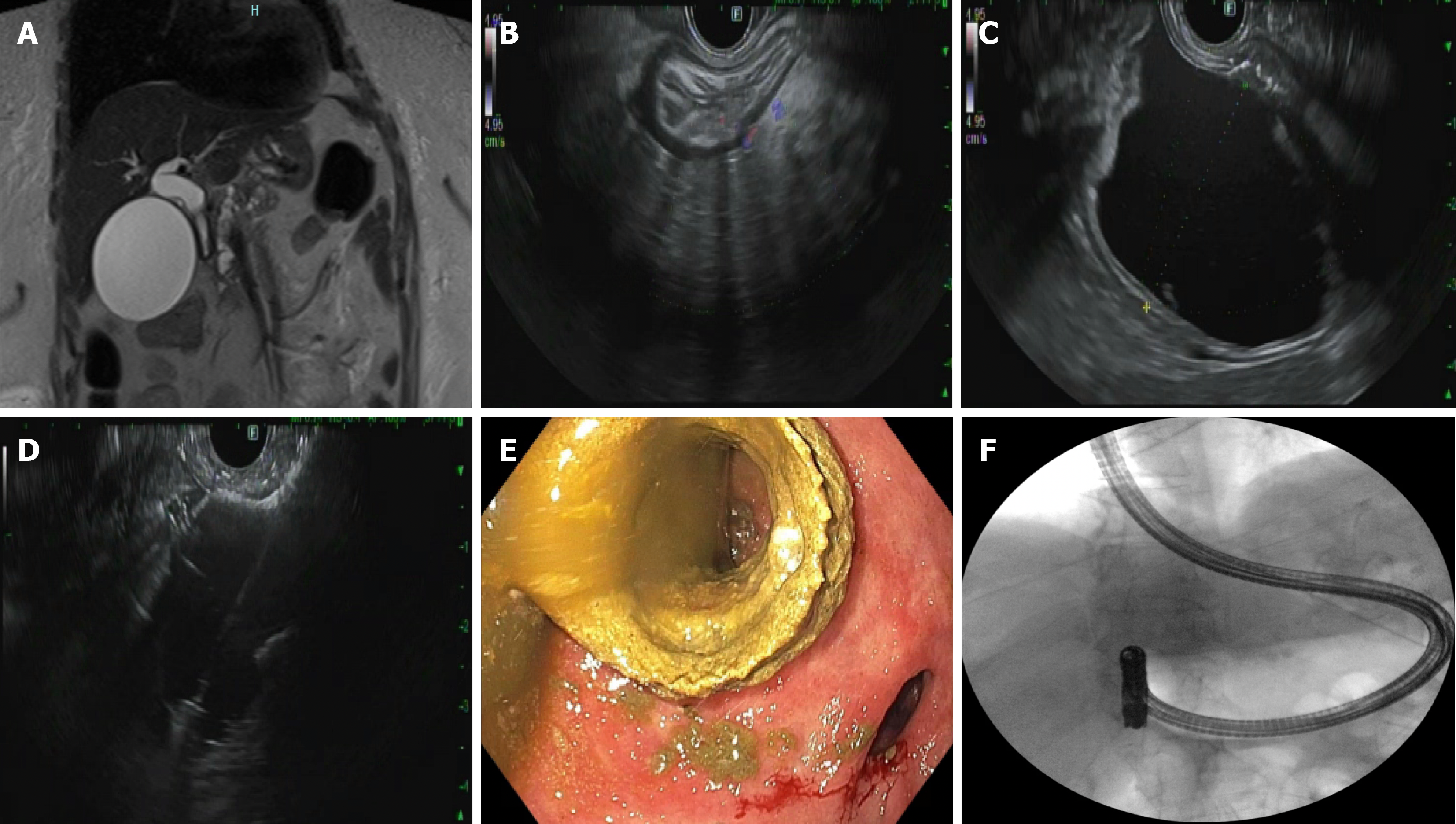
Figure 2 A 67-year-old female with a history of Roux-en-Y gastric bypass presented with abdominal pain attributed to chronic calcific pancreatitis, endoscopic ultrasound-directed transgastric endoscopic retrograde cholangiopancreatography was performed to facilitate endoscopic therapy.
A: Magnetic resonance imaging demonstrated a pancreatic duct stricture with upstream ductal dilation; B: Endoscopic ultrasound visualization of the excluded (remnant) stomach from the gastric pouch; C: The excluded stomach was distended under endosonographic guidance using a mixture of contrast and water; D: A 20-mm lumen-apposing metal stent was deployed to create a gastrogastric fistula; E: Endoscopic view confirming appropriate deployment of the proximal flange within the gastric pouch; F: A side-viewing duodenoscope was advanced through the stent into the remnant stomach to complete the endoscopic retrograde cholangiopancreatography.
- Citation: Saleh A, Yessin A, Salva W, Batarseh M, El Khoury M, Nehme F. Endoscopic ultrasound-guided gastrointestinal anastomosis: Evolving applications in advanced therapeutic endoscopy. World J Gastroenterol 2026; 32(39): 120546
- URL: https://www.wjgnet.com/1007-9327/full/v32/i39/120546.htm
- DOI: https://dx.doi.org/10.3748/wjg.120546