
















Copyright: ©Author(s) 2026.
World J Gastroenterol. Oct 14, 2026; 32(38): 121425
Published online Oct 14, 2026. doi: 10.3748/wjg.121425
Published online Oct 14, 2026. doi: 10.3748/wjg.121425
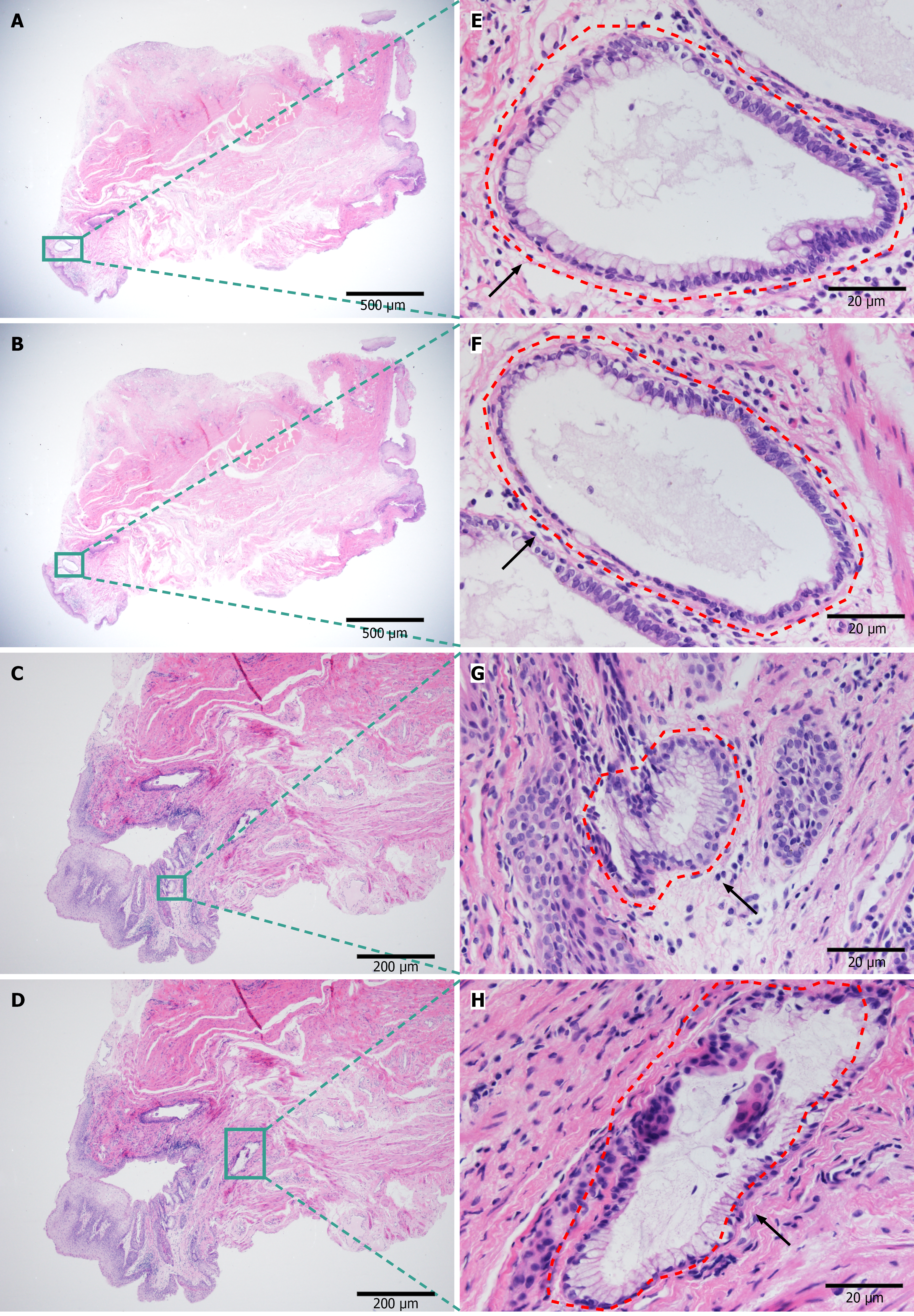
Figure 1 Histopathology of the internal-opening in cryptoglandular anal fistula (hematoxylin and eosin).
A-D: Low-power views of internal-opening tissue; E-H: Higher-power images of the boxed regions. Red dashed contours delineate gland-like structures. A 1-2 cell thick layer of spindle-shaped myoepithelial cells with goblet cells surrounding an anal gland, surrounded by florid chronic inflammatory infiltrates with fibroblast proliferation and collagen deposition. These features are consistent with a glandular origin of the fistula and were reviewed by a senior pathologist.
- Citation: Li TT, Li JN, Yang HW, Dou XY, Jiang L, Lai LX, Yu Q, Chen XY, Wang Y, Zhang XC, Ma HF, Song XB. Single-cell and bulk transcriptomics with machine learning decode B cell hub genes and diagnostic biomarkers in anal fistula. World J Gastroenterol 2026; 32(38): 121425
- URL: https://www.wjgnet.com/1007-9327/full/v32/i38/121425.htm
- DOI: https://dx.doi.org/10.3748/wjg.121425