
















Copyright: ©Author(s) 2026.
World J Gastroenterol. Oct 14, 2026; 32(38): 120967
Published online Oct 14, 2026. doi: 10.3748/wjg.120967
Published online Oct 14, 2026. doi: 10.3748/wjg.120967
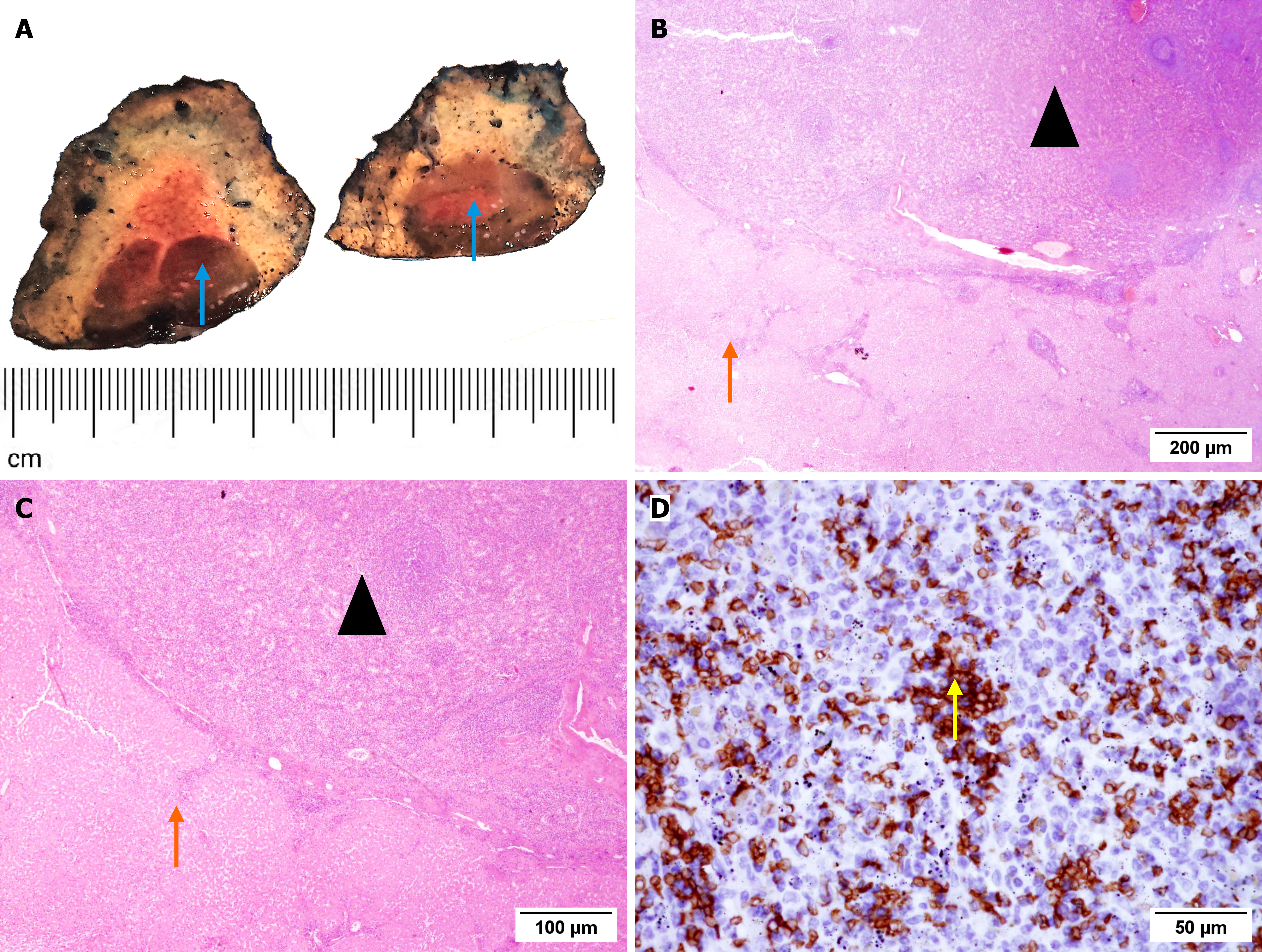
Figure 2 Histopathological analysis and immunohistochemical findings of the resected specimen.
A: Gross examination of the resected liver segment measuring 44 cm × 4.0 cm × 2.3 cm shows a dark red lesion (blue arrow) 1.6 cm in diameter, surrounded by a lighter rim; B and C: Hematoxylin and eosin staining [magnification × 10 (B) and × 20 (C)] demonstrates splenic tissue (triangle) surrounded by liver parenchyma (orange arrow); D: Immunohistochemical staining shows positivity for glycophorin C in erythroid cells (yellow arrow), magnification × 40.
- Citation: Sikiric M, Premec H, Mijic M, Sobocan N, Filipec Kanizaj T, Lovric E, Kavur L, Lalovac M. Diagnostic challenge of intrahepatic splenosis masquerading as hepatocellular carcinoma: A case report and review of literature. World J Gastroenterol 2026; 32(38): 120967
- URL: https://www.wjgnet.com/1007-9327/full/v32/i38/120967.htm
- DOI: https://dx.doi.org/10.3748/wjg.120967