
















Copyright: ©Author(s) 2026.
World J Gastroenterol. Oct 7, 2026; 32(37): 120860
Published online Oct 7, 2026. doi: 10.3748/wjg.120860
Published online Oct 7, 2026. doi: 10.3748/wjg.120860
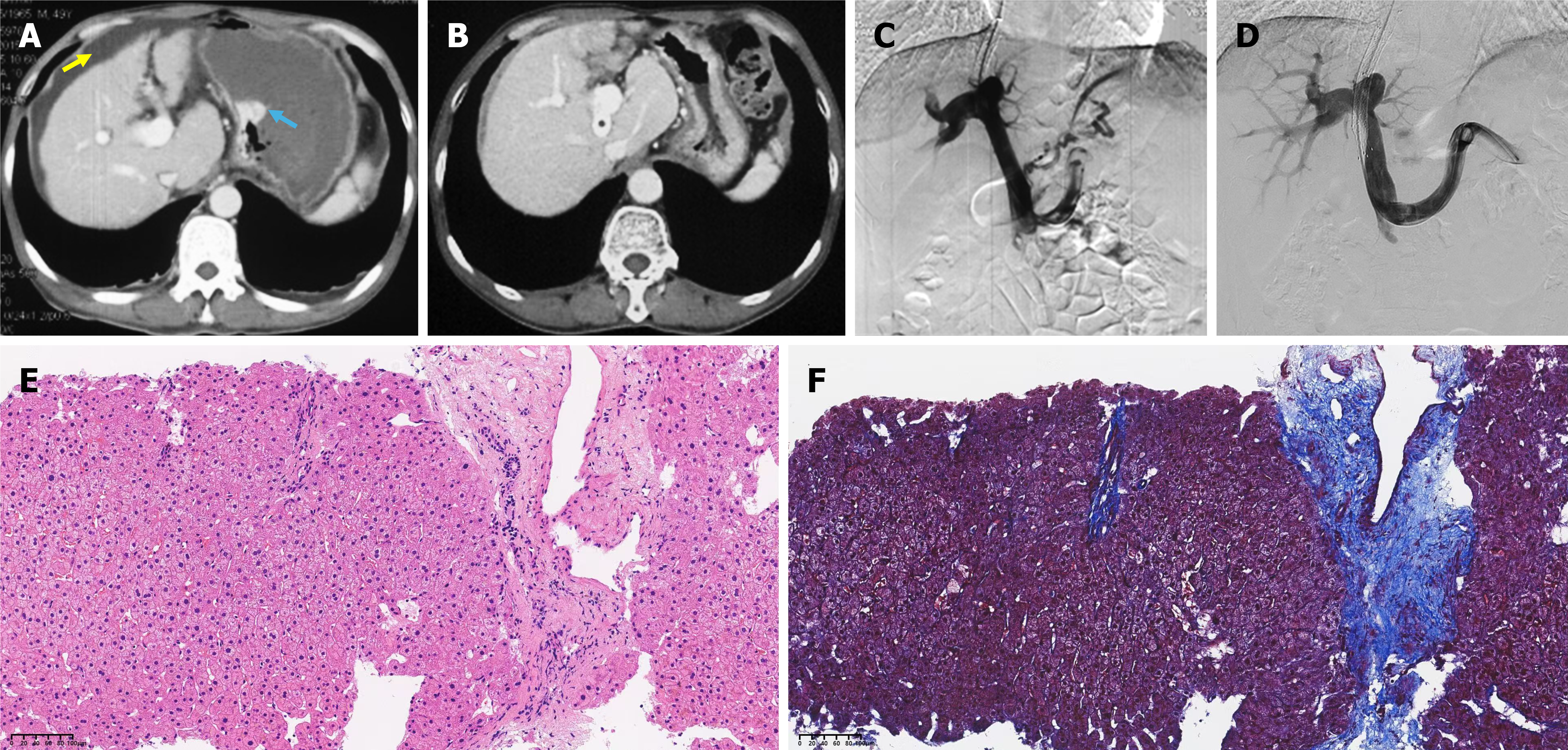
Figure 2 Imaging and histopathology illustrate portal pressure changes and hepatic fibrosis regression after transjugular intrahepatic portosystemic shunt.
A: Baseline contrast-enhanced computed tomography showing cirrhosis with a small-volume ascites (yellow arrow) and esophagogastric varices (blue arrow); B: Final follow-up computed tomography demonstrating resolution of ascites and varices despite occlusion of the transjugular intrahepatic portosystemic shunt (TIPS) stent; C: Direct portography performed 6 months after TIPS for recurrent gastrointestinal bleeding showing stent occlusion with recurrent varices; portal pressure gradient was 25 mmHg, and TIPS revision was performed; D: Direct portography 10 years after TIPS showing stent occlusion without varices; portal pressure gradient was 6 mmHg, and no intervention was undertaken; E: Hematoxylin and eosin-stained liver tissue shows hepatocytes arranged in cords with preserved sinusoidal architecture. Interlobular septa are maintained with residual fibrous septa; there are no histologic features of cirrhosis or chronic inflammation; F: Masson’s trichrome staining reveals branched fibrous septa with expansion of portal fibrous tissue, without definite nodular remodeling or classic cirrhotic features. The fibrosis stage is Ishak 3.
- Citation: Tie J, Yuan XL, Niu J, Xu J, Shi YQ. Transjugular intrahepatic portosystemic shunt combined with viral suppression reverses decompensated cirrhosis: Two case reports. World J Gastroenterol 2026; 32(37): 120860
- URL: https://www.wjgnet.com/1007-9327/full/v32/i37/120860.htm
- DOI: https://dx.doi.org/10.3748/wjg.120860