
















Copyright: ©Author(s) 2026.
World J Gastroenterol. Sep 28, 2026; 32(36): 120568
Published online Sep 28, 2026. doi: 10.3748/wjg.120568
Published online Sep 28, 2026. doi: 10.3748/wjg.120568
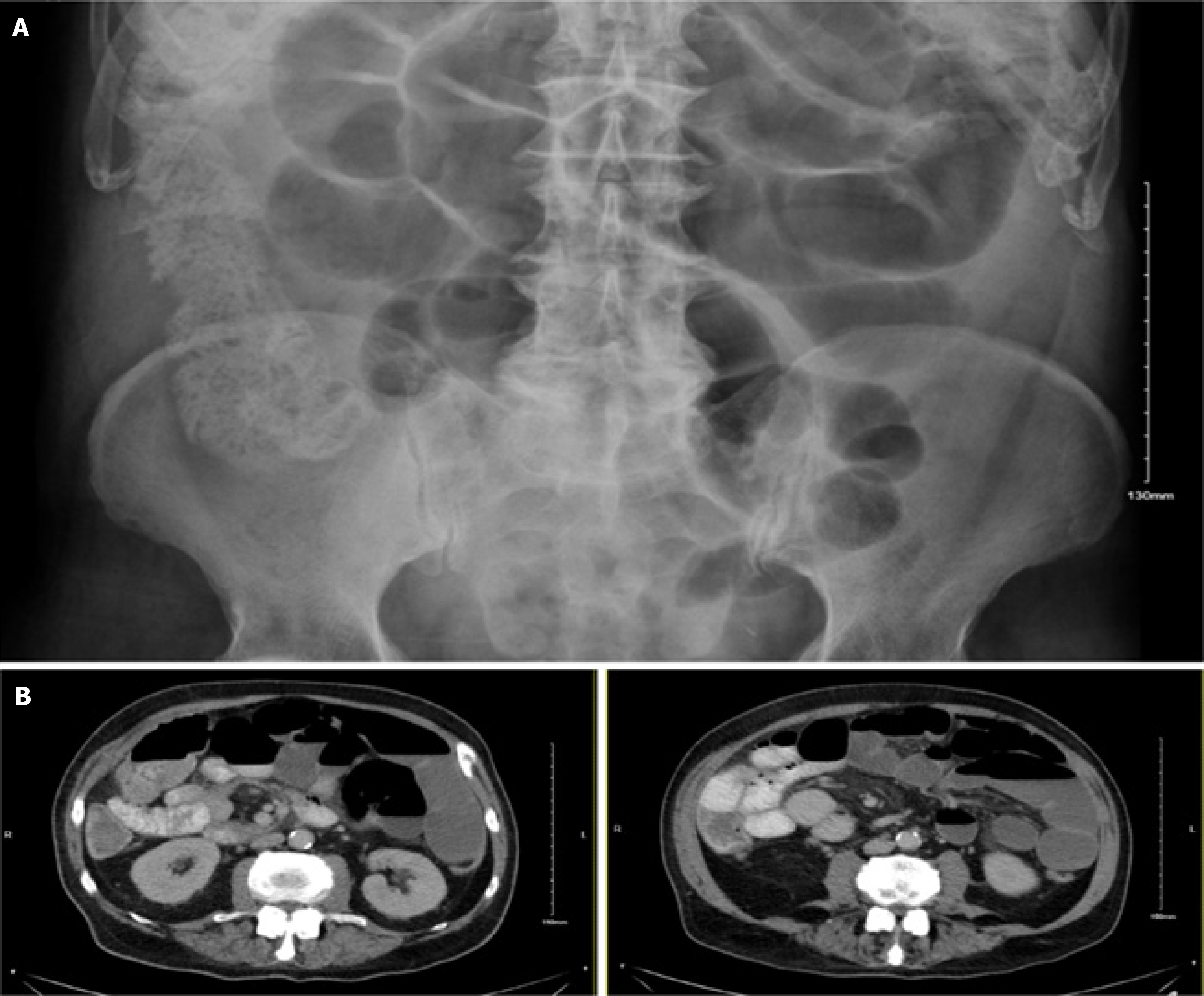
Figure 1 Abdominal radiography and computed tomography findings.
A: Abdominal radiograph showing a bowel gas pattern within normal limits, with evidence of fecal loading; B: Contrast-enhanced computed tomography of the abdomen and pelvis demonstrating mild dilatation of the jejunum with fluid-filled loops and associated fecalization of the ileum. The distal ileum and ileocaecal valve are of normal caliber. The large bowel is relatively decompressed, with no intra-abdominal free fluid or air.
- Citation: Kharief M, O’Byrne R, William K, Hasabarsoul MA, Tuohy O, Morcos A. Anti-glutamic acid decarboxylase autoimmunity presenting as chronic intestinal pseudo-obstruction with dysautonomia and late-onset autoimmune diabetes: A case report. World J Gastroenterol 2026; 32(36): 120568
- URL: https://www.wjgnet.com/1007-9327/full/v32/i36/120568.htm
- DOI: https://dx.doi.org/10.3748/wjg.120568