
















Copyright: ©Author(s) 2026.
World J Gastroenterol. Sep 14, 2026; 32(34): 120518
Published online Sep 14, 2026. doi: 10.3748/wjg.v32.i34.120518
Published online Sep 14, 2026. doi: 10.3748/wjg.v32.i34.120518
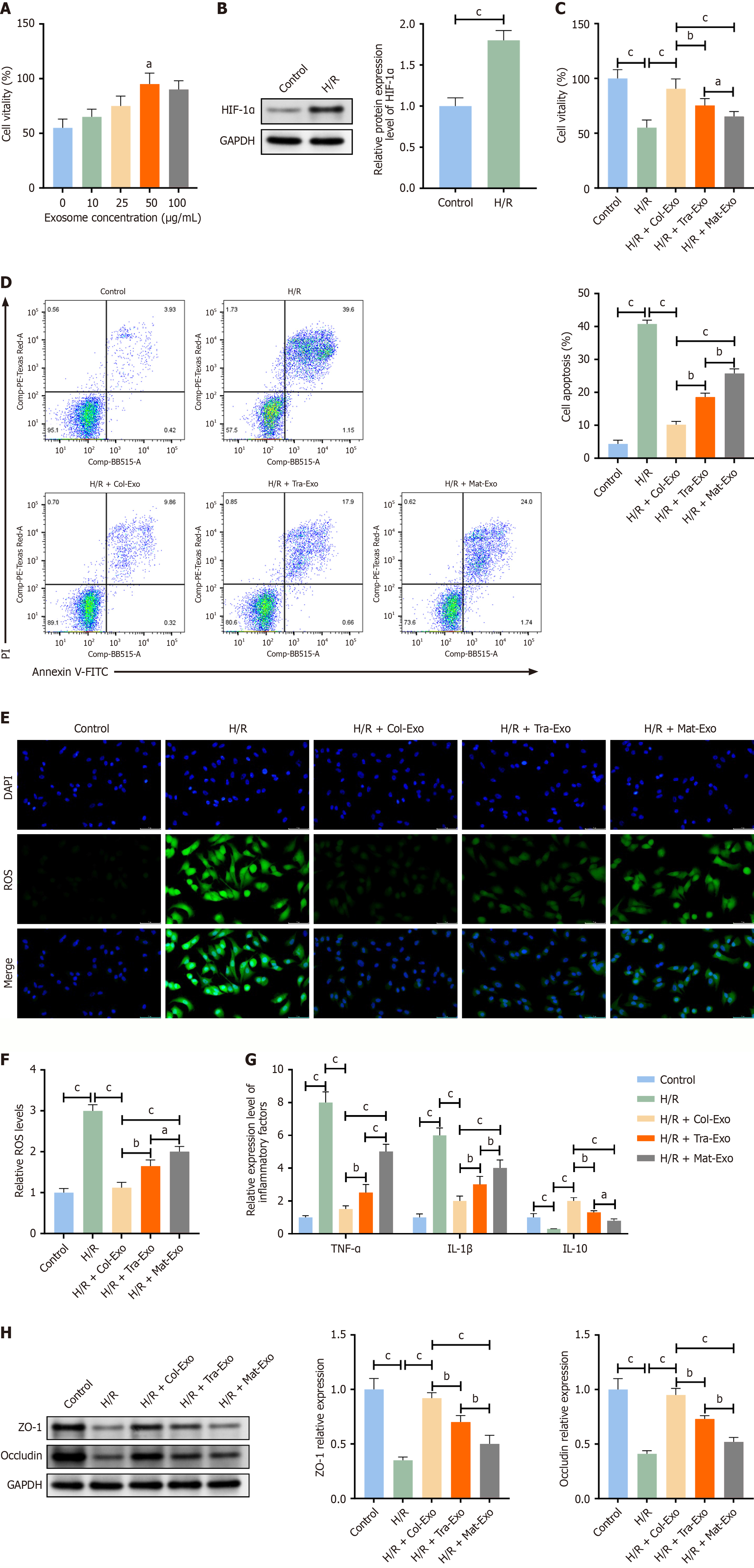
Figure 3 Protective effects of stage - specific exosomes derived from human milk on Caco-2 cells subjected to hypoxia/reoxygenation injury.
A: Cell viability assessed by CCK-8 assay to determine the optimal exosome concentration; B: Western blot analysis of hypoxia-inducible factor-1α expression; C: Cell viability under hypoxia/reoxygenation injury with or without exosome treatment; D: Apoptosis rates detected by Annexin V-FITC/PI staining; E: Representative fluorescence images of intracellular reactive oxygen species levels (scale bar = 50 μm; × 400); F: Quantification of reactive oxygen species fluorescence intensity; G: Levels of tumor necrosis factor-α, interleukin-1β, and interleukin-10 in cell supernatants; H: Western blot analysis of tight junction proteins zonula occludens-1 and occludin. aP < 0.05, bP < 0.01, cP < 0.001. HIF-1α: Hypoxia-inducible factor-1α; IL: Interleukin; H/R: Hypoxia/reoxygenation; ROS: Reactive oxygen species; ZO-1: Zonula occludens-1.
- Citation: Yang G, Liu L. Lactation stage-dependent protective effects of human milk exosomes against intestinal ischemia-reperfusion injury via lactoferrin/lysozyme-mediated Nrf2/HO-1 and NF-κB pathways. World J Gastroenterol 2026; 32(34): 120518
- URL: https://www.wjgnet.com/1007-9327/full/v32/i34/120518.htm
- DOI: https://dx.doi.org/10.3748/wjg.v32.i34.120518