
















Copyright: ©Author(s) 2026.
World J Gastroenterol. Sep 14, 2026; 32(34): 120518
Published online Sep 14, 2026. doi: 10.3748/wjg.v32.i34.120518
Published online Sep 14, 2026. doi: 10.3748/wjg.v32.i34.120518
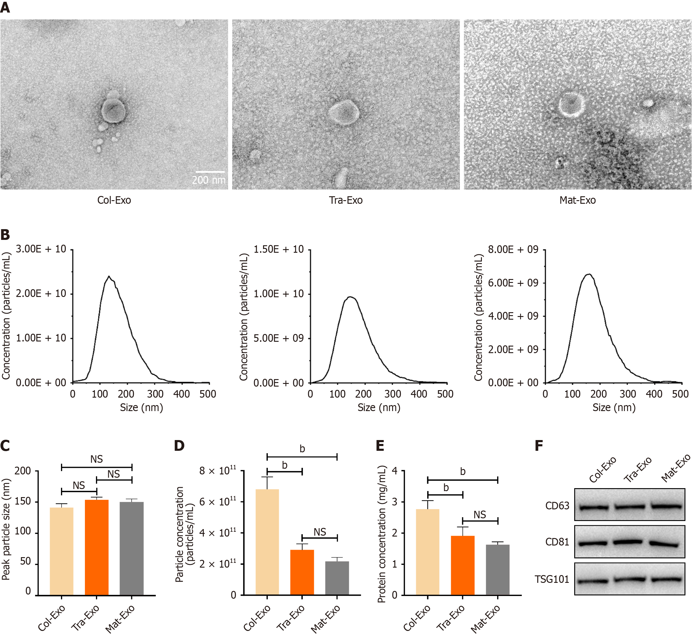
Figure 2 Identification of extracellular vesicles derived from breast milk at different lactation stages.
A: Representative transmission electron microscopy images showing Col-Exo, Tra-Exo, and Mat-Exo (scale bar = 200 nm); B: Nanoparticle tracking analysis showed the size distribution characteristics of extracellular vesicles at different stages; C: Peak particle size; D: Particle concentration; E: Protein concentration; F: Western blot analysis of extracellular vesicle markers CD63, CD81, and tumor susceptibility gene 101. bP < 0.01. NS: Not significant; TSG101: Tumor susceptibility gene 101.
- Citation: Yang G, Liu L. Lactation stage-dependent protective effects of human milk exosomes against intestinal ischemia-reperfusion injury via lactoferrin/lysozyme-mediated Nrf2/HO-1 and NF-κB pathways. World J Gastroenterol 2026; 32(34): 120518
- URL: https://www.wjgnet.com/1007-9327/full/v32/i34/120518.htm
- DOI: https://dx.doi.org/10.3748/wjg.v32.i34.120518