
















Copyright: ©Author(s) 2026.
World J Gastroenterol. Sep 14, 2026; 32(34): 119467
Published online Sep 14, 2026. doi: 10.3748/wjg.119467
Published online Sep 14, 2026. doi: 10.3748/wjg.119467
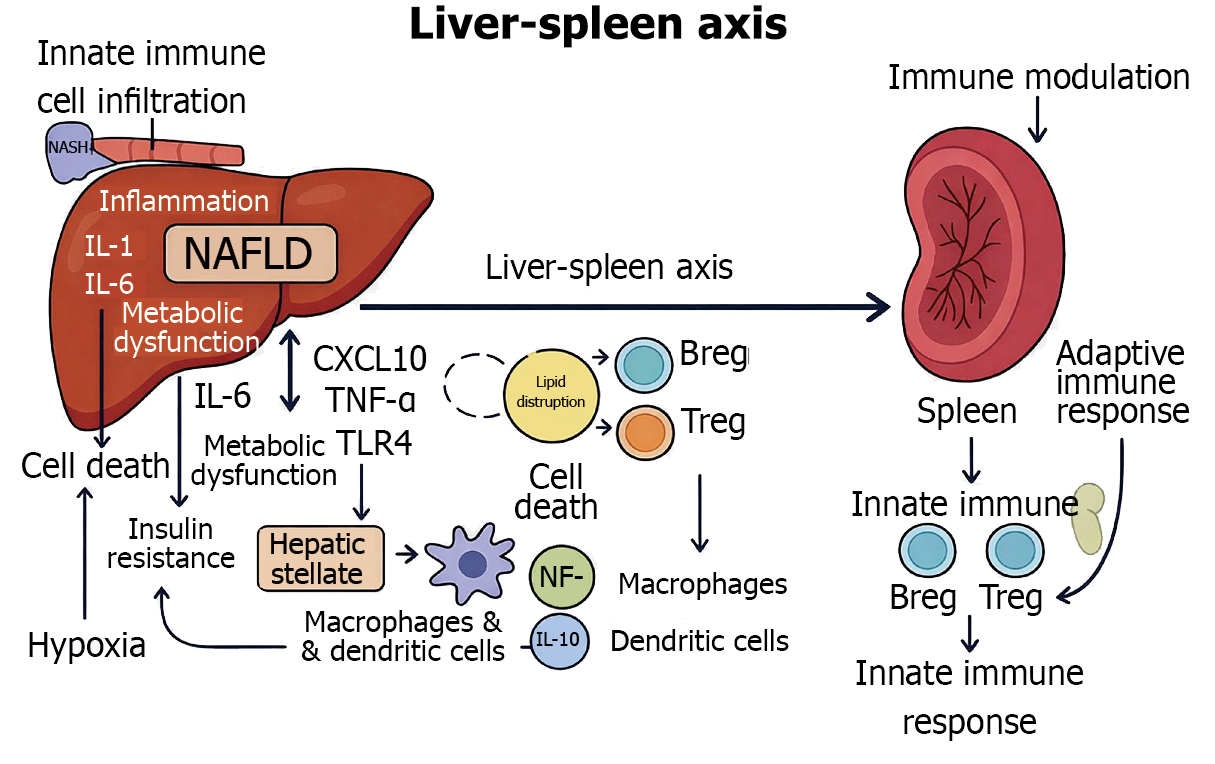
Figure 1 Crosstalk between the spleen and liver.
The liver-spleen axis is governed by a dynamic bidirectional immune-cell trafficking network in which splenic monocytes, natural killer (NK) cells, and lymphocytes are mobilized via sphingosine-1-phosphate (S1P)/S1P receptor 1-driven egress, guided through the portal circulation by C-X-C motif chemokine ligand 9/10 (CXCL9/10) and CXCL12 chemokine gradients, arrested on liver sinusoidal endothelial cells through selectin-P-selectin glycoprotein ligand-1 and intercellular adhesion molecule-1/Lymphocyte function-associated antigen-1 integrin-mediated firm adhesion, and conditioned within the hepatic microenvironment by reciprocal cytokine signals including interleukin (IL-6)/Janus kinase/signal transducer and activator of transcription 3, interferon gamma, transforming growth factor-beta/SMAD, and tumor necrosis factor alpha, while the liver in turn reprograms splenic immune tone through portal delivery of lipopolysaccharides/Toll-like receptor 4 (TLR4) agonists, bile acids, damage-associated molecular patterns-driven NOD-like receptor pyrin domain-containing protein 3/IL-1β, hepatokines such as fibroblast growth factor 21 and hepatocyte growth factor, and extracellular vesicle-encapsulated microRNA cargo, with the entire axis held in homeostatic balance by regulatory checkpoints including programmed cell death protein 1/programmed death-ligand 1 on liver sinusoidal endothelial cells, T-cell immunoglobulin and mucin domain-containing protein 3/galectin-9-mediated NK and T-cell exhaustion, cytotoxic T-lymphocyte associated protein 4/cluster of differentiation 80 (CD80)/CD86 regulatory T cell (Treg) suppression, and indoleamine-driven tryptophan depletion that collectively sustain hepatic immune tolerance. Cellular players: Kupffer cells, hepatic stellate cells, Tregs, regulatory B cells (Bregs), macrophages, and dendritic cells. Immune signaling: Tumor necrosis factor alpha (TNF-α), IL-6, IL-10, CXCL10, nuclear factor kappa B (NF-κB), TLR4; nonalcoholic steatohepatitis (NASH). Pathological progression: From steatosis to NASH, including fibrosis and hypoxia. Metabolic dysfunction: Lipid disruption, insulin resistance, de novo lipogenesis.
- Citation: Tarantino G, Citro V. Liver-spleen axis: Deciphering a crucial crosstalk in non-alcoholic fatty liver disease progression and therapeutic implications. World J Gastroenterol 2026; 32(34): 119467
- URL: https://www.wjgnet.com/1007-9327/full/v32/i34/119467.htm
- DOI: https://dx.doi.org/10.3748/wjg.119467