
















Copyright: ©Author(s) 2026.
World J Gastroenterol. Aug 28, 2026; 32(32): 120450
Published online Aug 28, 2026. doi: 10.3748/wjg.120450
Published online Aug 28, 2026. doi: 10.3748/wjg.120450
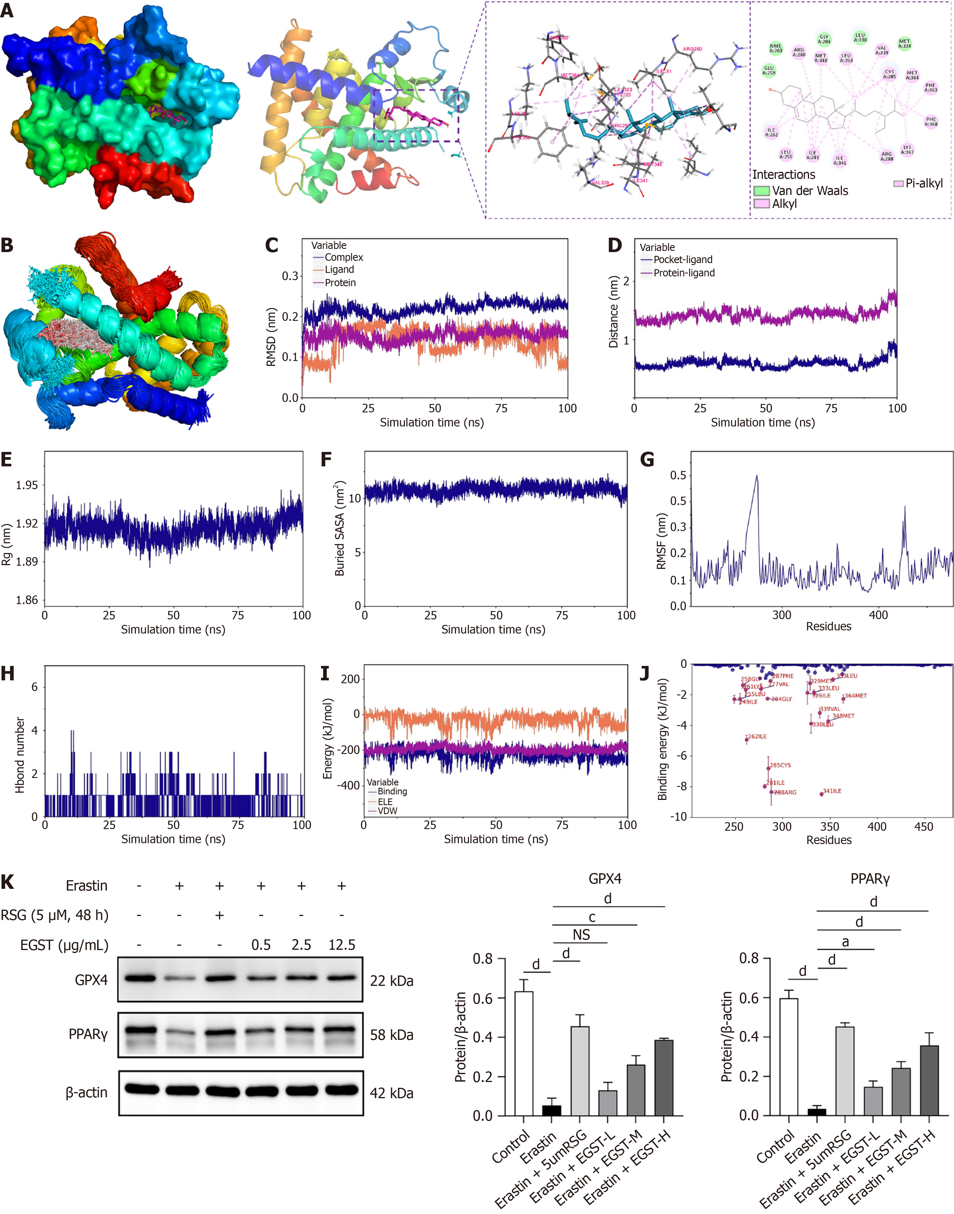
Figure 6 Molecular dynamics simulation and western blot analysis of PPARγ and glutathione peroxidase 4.
A: Molecular docking mode of beta-sitosterol with peroxisome proliferator-activated receptor gamma (PPARγ); B: 3D representation of beta-sitosterol; C: Root means square deviation between beta-sitosterol and PPARγ; D: Distance analysis between beta-sitosterol and PPARγ; E: Radius of gyration of beta-sitosterol and PPARγ; F: Solvent-accessible surface area analysis; G: Root means square fluctuation analysis of beta-sitosterol vs PPARγ; H: Hydrogen bond analysis between RhoA and AT-1; I: Binding energy analysis; J: Amino acid binding energy contribution ranking; K: Western blot analysis of PPARγ and glutathione peroxidase 4 protein expression following extracts from Gleditsia sinensis thorns and rosiglitazone treatment. Data are presented as mean ± SD (n = 3). aP < 0.05, cP < 0.001, and dP < 0.0001. DSS: Dextran sulfate sodium; EGST: Extracts from Gleditsia sinensis thorns; NS: Not significant; RSG: Rosiglitazone; GPX4: Glutathione peroxidase 4; PPARγ: Peroxisome proliferator-activated receptor gamma.
- Citation: Zhuo ZY, Guo XY, Liu XM, Shang MM, Xue S, Peng L, Guo ZG. Extracts of Gleditsia sinensis Lam. thorn ameliorates ulcerative colitis through PPARγ/GPX4 pathway-mediated inhibition of enterocyte ferroptosis. World J Gastroenterol 2026; 32(32): 120450
- URL: https://www.wjgnet.com/1007-9327/full/v32/i32/120450.htm
- DOI: https://dx.doi.org/10.3748/wjg.120450