
















Copyright: ©Author(s) 2026.
World J Gastroenterol. Aug 28, 2026; 32(32): 120382
Published online Aug 28, 2026. doi: 10.3748/wjg.120382
Published online Aug 28, 2026. doi: 10.3748/wjg.120382
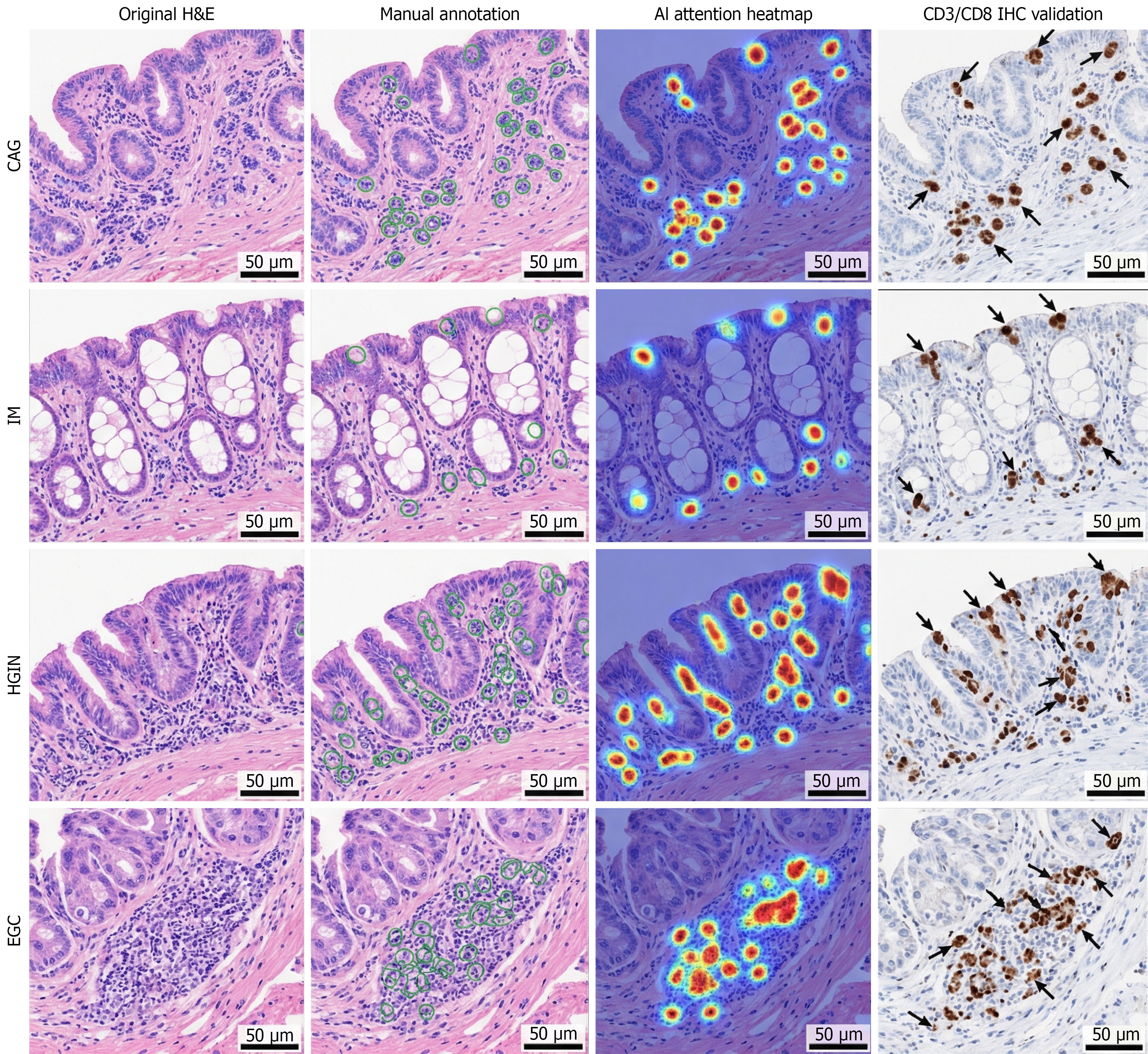
Figure 4 Model interpretability analysis and immunohistochemistry validation of tumour-infiltrating lymphocyte recognition.
Representative images (scale bar = 50 μm) of chronic atrophic gastritis, intestinal metaplasia, high-grade intraepithelial neoplasia and early gastric cancer compared with original hematoxylin and eosin staining, pathologists’ double-blind manual annotation, artificial intelligence (AI) attention heatmaps and CD3/CD8 immunohistochemistry (IHC) validation. Red regions in heatmaps indicate the model’s focus on tumour-infiltrating lymphocyte (TIL) nuclear regions, avoiding interference from goblet cells (IM). AI-recognized epithelial TIL regions show 100% positive concordance with CD3/CD8 IHC staining (black arrows), confirming that there are no false positives. The attention regions of the model are highly consistent with the manual annotations (IoU = 0.89; Dice coefficient = 0.94). CAG: Chronic atrophic gastritis; IM: Intestinal metaplasia; HGIN: High-grade intraepithelial neoplasia; EGC: Early gastric cancer; IHC: Immunohistochemistry; AI: Artificial intelligence; H/E: Hematoxylin and eosin.
- Citation: Fan Y, Wang SN, Jiang B, Li YY, Zhu CY, Liao XH, Zhang FS, Wang YK. Automatic recognition of tumour-infiltrating lymphocytes in pathological biopsy images of the gastric mucosa. World J Gastroenterol 2026; 32(32): 120382
- URL: https://www.wjgnet.com/1007-9327/full/v32/i32/120382.htm
- DOI: https://dx.doi.org/10.3748/wjg.120382