
















Copyright: ©Author(s) 2026.
World J Gastroenterol. Aug 28, 2026; 32(32): 120382
Published online Aug 28, 2026. doi: 10.3748/wjg.120382
Published online Aug 28, 2026. doi: 10.3748/wjg.120382
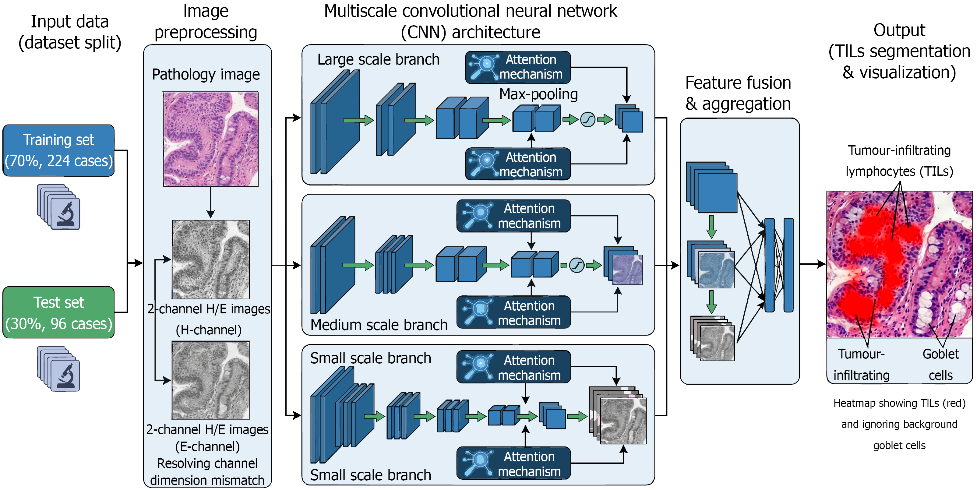
Figure 2 Schematic workflow and multiscale convolutional neural network architecture for tumour-infiltrating lymphocyte recognition in gastric mucosa.
The pipeline consists of four primary stages: (1) Dataset preparation: The study cohort was divided into a training set and an independent test set; (2) Image preprocessing: Original hematoxylin and eosin images were decomposed into 2-channel (H and E) components via color deconvolution to resolve channel dimension mismatch and enhance staining robustness; (3) Model core: A multiscale convolutional neural network architecture featuring parallel branches and attention mechanisms was employed to extract multi-resolution features; and (4) Output and visualization: The model generates attention heatmaps that precisely highlight tumour-infiltrating lymphocyte nuclear regions (red) while effectively suppressing interference from background structures such as goblet cells. H/E: Hematoxylin and eosin.
- Citation: Fan Y, Wang SN, Jiang B, Li YY, Zhu CY, Liao XH, Zhang FS, Wang YK. Automatic recognition of tumour-infiltrating lymphocytes in pathological biopsy images of the gastric mucosa. World J Gastroenterol 2026; 32(32): 120382
- URL: https://www.wjgnet.com/1007-9327/full/v32/i32/120382.htm
- DOI: https://dx.doi.org/10.3748/wjg.120382