
















Copyright: ©Author(s) 2026.
World J Gastroenterol. Aug 28, 2026; 32(32): 120382
Published online Aug 28, 2026. doi: 10.3748/wjg.120382
Published online Aug 28, 2026. doi: 10.3748/wjg.120382
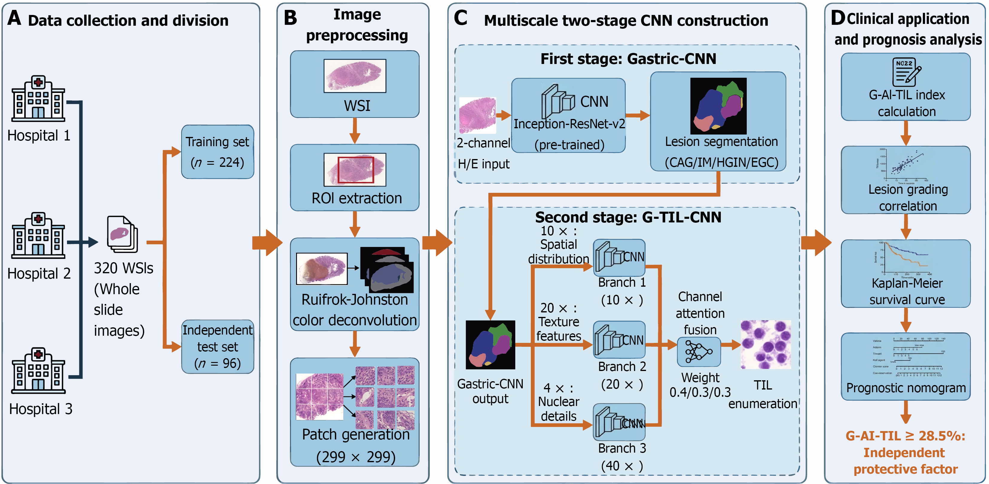
Figure 1 Overall workflow of the multiscale two-stage convolutional neural network model for automatic recognition of gastric mucosal tumour-infiltrating lymphocytes and prognostic analysis.
This workflow shows four core stages of automated tumour-infiltrating lymphocyte (TIL) analysis and prognostic evaluation in gastric mucosal biopsy images. A: A total of 320 whole slide images (WSIs) from three centres were divided into training and in
- Citation: Fan Y, Wang SN, Jiang B, Li YY, Zhu CY, Liao XH, Zhang FS, Wang YK. Automatic recognition of tumour-infiltrating lymphocytes in pathological biopsy images of the gastric mucosa. World J Gastroenterol 2026; 32(32): 120382
- URL: https://www.wjgnet.com/1007-9327/full/v32/i32/120382.htm
- DOI: https://dx.doi.org/10.3748/wjg.120382