
















Copyright: ©Author(s) 2026.
World J Gastroenterol. Aug 21, 2026; 32(31): 122449
Published online Aug 21, 2026. doi: 10.3748/wjg.122449
Published online Aug 21, 2026. doi: 10.3748/wjg.122449
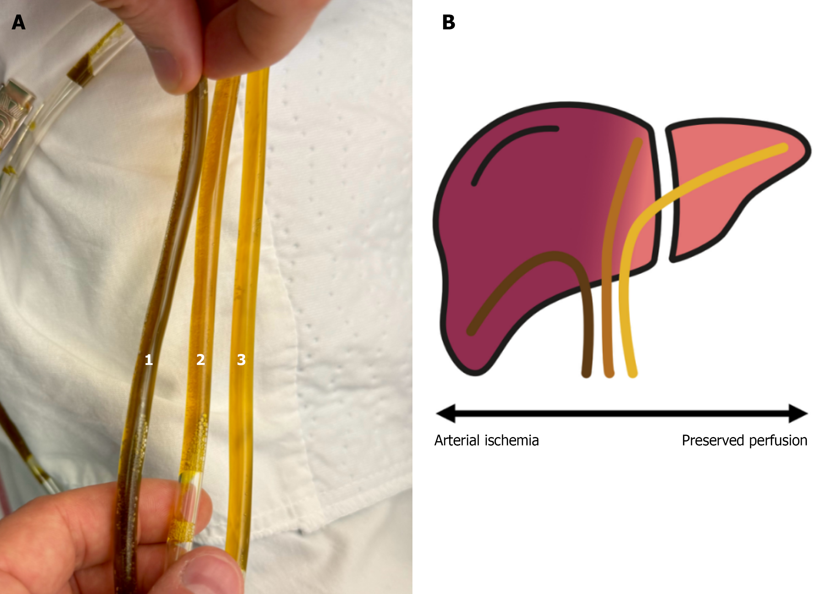
Figure 4 The “biliary sunrise”.
(Figure 4B was created by Adrian Stelzl using a professional graphic illustration software - Affinity Designer). A: Extracted bile demonstrating a gradient of biliary pigmentation from more ischemic to less ischemic liver regions. From left to right: 1: Right posterolateral duct; 2: Right anteromedial duct; 3: Left bile duct; B: Schematic illustration of Neuhaus drain positioning within the partially ischemic liver.
- Citation: Stelzl A, El-Mahrouk M, Stiegler P, Wagner M, Sucher R. Biliary sunrise - bile color as visible sign for hepatic ischemia after iatrogenic partial liver de-arterialization: A case report. World J Gastroenterol 2026; 32(31): 122449
- URL: https://www.wjgnet.com/1007-9327/full/v32/i31/122449.htm
- DOI: https://dx.doi.org/10.3748/wjg.122449