
















Copyright: ©Author(s) 2026.
World J Gastroenterol. Aug 21, 2026; 32(31): 118472
Published online Aug 21, 2026. doi: 10.3748/wjg.118472
Published online Aug 21, 2026. doi: 10.3748/wjg.118472
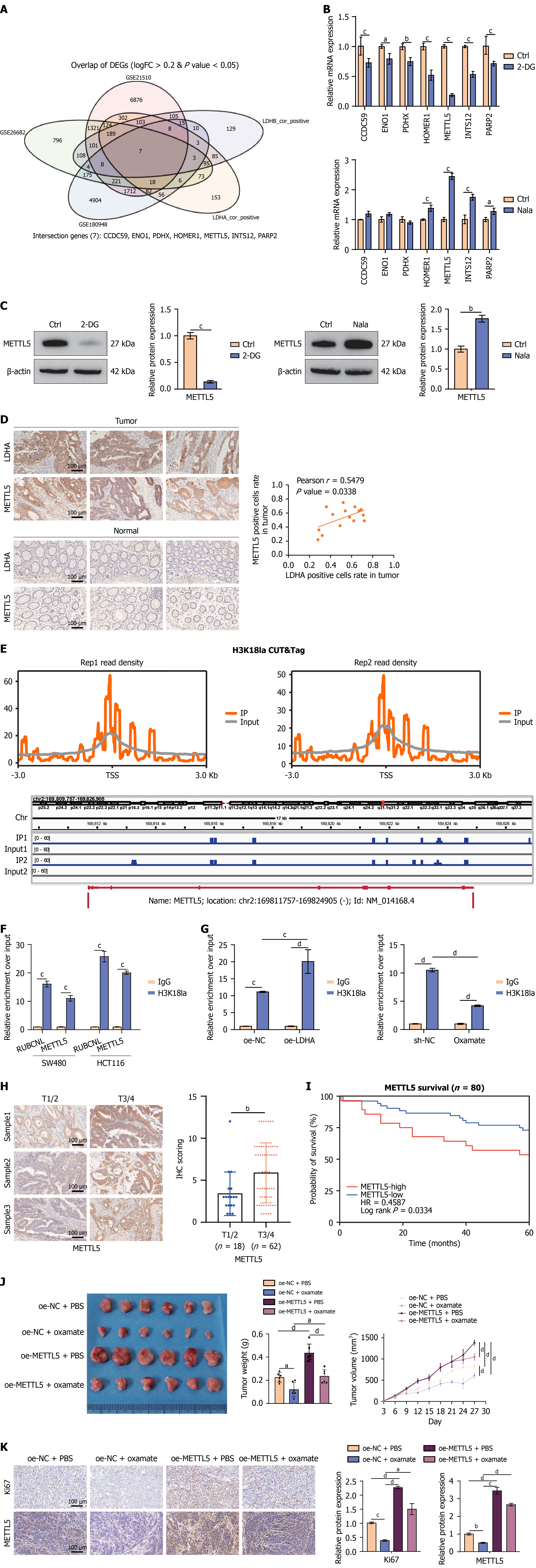
Figure 2 Lactate and H3K18 La promote METTL5 transcription.
A: Venn diagram illustrating overlapping genes among gene sets highly expressed in colorectal cancer (CRC) tissues (GSE21510, GSE26692, and GSE180948) and gene sets positively correlated with LDHA or LDHB expression (LDHA_cor_positive, LDHB_cor_positive); B: SW480 cells treated with 5 mmol/L 2-DG or 5 mmol/L sodium lactate (Nala) for 24 hours. Quantitative polymerase chain reaction (qPCR) to detect the mRNA expression levels of CCDC59, ENO1, PDHX, HOMER1, METTL5, INTS12, and PARP2; C: SW480 cells treated with 5 mmol/L 2-DG or 5 mmol/L Nala for 24 hours. Western blotting to detect METTL5 protein expression; β-actin served as the internal reference; D: Immunohistochemistry (IHC) of METTL5 and LDHA protein expression in 15 randomly selected CRC tissues (T) and adjacent non-tumor tissues (N) (left panel). Positive cell proportion and correlation between METTL5 and LDHA expression were analyzed; E: SW480 cells treated with 5 mmol/L Nala for 24 hours, followed by the cleavage under targets and tagmentation assay using H3K18 La antibody and IgG control antibody. The upper panel depicts the genome-wide enrichment of H3K18 La near transcription start sites; the lower panel displays the sequencing signal peaks of H3K18 La and IgG in the chromosomal region of METTL5; F: Chromatin immunoprecipitation in SW480 and HCT116 cells using H3K18 La antibody and IgG, respectively. qPCR to detect the enrichment level of H3K18 La in the region near the METTL5 promoter; G: SW480 cells transfected with LDHA-overexpressing plasmid (left) or treated with 10 mmol/L oxamate for 24 hours (right). A subsequent chromatin immunoprecipitation assay was conducted, and qPCR was used to analyze the enrichment of H3K18 La in the METTL5 promoter region; H: IHC quantification of METTL5 expression in 80 CRC tissue samples (left panel, scale bar: 100 μm). The Allred scoring system to quantify METTL5 expression level (right panel); I: Survival analysis of the 80 patients. Based on the Allred score of METTL5, patients were divided into low-expression (score ≤ 2, n = 23) and high-expression (score ≥ 3, n = 57) groups. Differences in overall survival between the two groups were compared; J: SW480 cells stably transfected with empty vector plasmid or METTL5-overexpressing plasmid (1 × 10⁶ cells/mouse) were subcutaneously inoculated into BALB/c nude mice to establish a cell-derived xenograft model. Tumor volume was measured every 3 days starting from day 3 post-inoculation. On day 6 post-inoculation, mice were administered phosphate-buffered saline (control group) or oxamate (750 mg/kg, intraperitoneal injection daily). Mice were euthanized on day 27 post-inoculation; tumors were dissected and photographed (left), weighed for statistical analysis (middle, n = 6), and tumor growth curves were plotted (right); K: IHC to detect Ki67 and METTL5 expression in xenograft tumors from different treatment groups (as in J), followed by quantitative analysis. PBS: Phosphate-buffered saline. aP < 0.05, bP < 0.01, cP < 0.001, and dP < 0.0001.
- Citation: Wang XP, Liu C, Zhang HW, Zhai JM, Zhu JX, Sun GD, An YH, Yang HJ, Jing DS, Liu DC. H3K18 La-METTL5-CCT2 signaling axis promotes colorectal cancer progression by inhibiting pyroptosis. World J Gastroenterol 2026; 32(31): 118472
- URL: https://www.wjgnet.com/1007-9327/full/v32/i31/118472.htm
- DOI: https://dx.doi.org/10.3748/wjg.118472