
















Copyright: ©Author(s) 2026.
World J Gastroenterol. Aug 21, 2026; 32(31): 118472
Published online Aug 21, 2026. doi: 10.3748/wjg.118472
Published online Aug 21, 2026. doi: 10.3748/wjg.118472
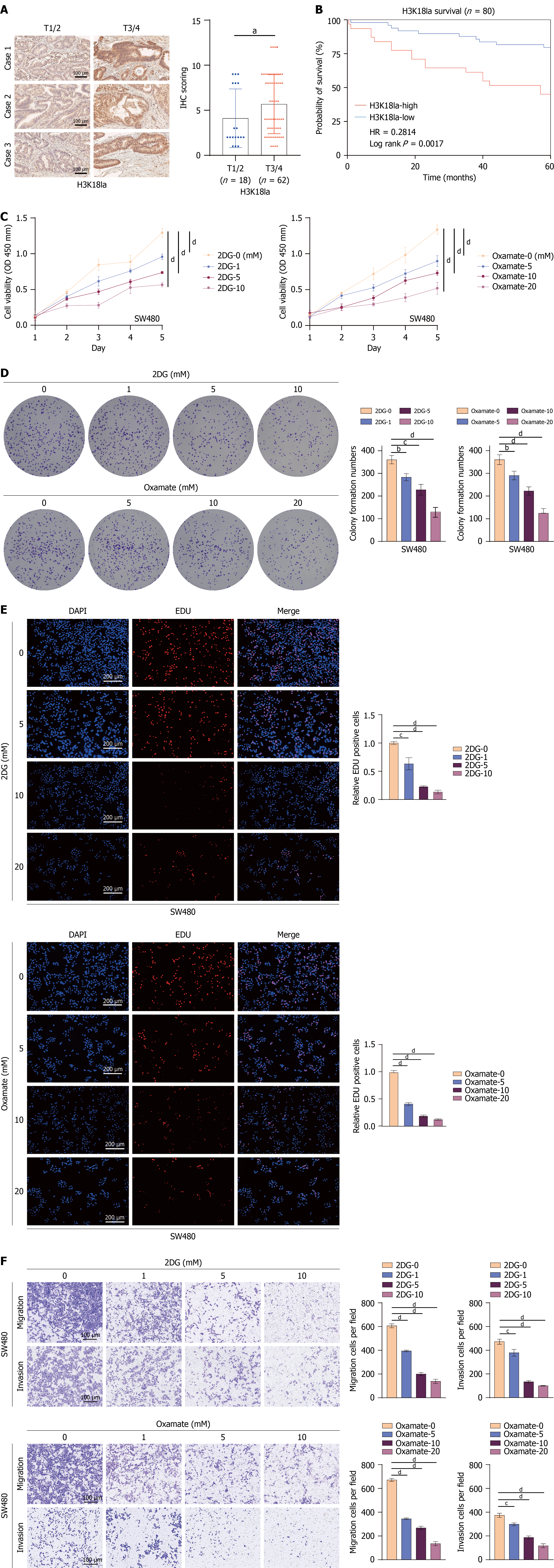
Figure 1 Lactylation is closely associated with the prognosis of patients with colorectal cancer.
A: Immunohistochemistry detection of H3K18 La in 80 colorectal cancer (CRC) tissue samples, including 18 cases at stage T1/2 and 62 cases at stage T3/4 (left panel, scale bar: 100 μm). H3K18 La expression was quantified using the Allred scoring system (right). Stage T1/2: In situ CRC without lymph node or distant metastasis; stage T3/4: Advanced CRC with lymph node or distant metastasis; B: Survival analysis of the 80 patients. Patients were divided into low-expression (Allred score ≤ 2, n = 24) and high-expression (score ≥ 3, n = 56) based on H3K18 La levels; C: SW480 cells (1 × 10⁴ cells/well) cultured with different concentrations of 2-DG or oxamate for 5 days. Cell viability was detected using the cell counting kit-8 for 5 consecutive days, and cell growth curves were plotted; D: SW480 cells (5 × 10² cells/well) cultured with different concentrations of 2-DG or oxamate for 14 days. Subsequently, crystal violet staining was performed to visualize the formation of cell colonies, and the number of monoclonal cell clusters was counted and subjected to quantitative analysis; E: SW480 cells (5 × 10³ cells/well) cultured with different concentrations of 2-DG or oxamate for 24 hours and incubated with EdU for an additional 2 hours. The number of EdU-positive cells was detected by fluorescence microscopy and subjected to quantitative comparison; F: SW480 cells (1.5 × 10⁴ cells/chamber) seeded in Transwell insert or Matrigel-coated Transwell insert with medium containing 2-DG or oxamate. After 48 hours of culture, cells were stained with crystal violet (scale bar: 100 μm). aP < 0.05, bP < 0.01, cP < 0.001, and dP < 0.0001.
- Citation: Wang XP, Liu C, Zhang HW, Zhai JM, Zhu JX, Sun GD, An YH, Yang HJ, Jing DS, Liu DC. H3K18 La-METTL5-CCT2 signaling axis promotes colorectal cancer progression by inhibiting pyroptosis. World J Gastroenterol 2026; 32(31): 118472
- URL: https://www.wjgnet.com/1007-9327/full/v32/i31/118472.htm
- DOI: https://dx.doi.org/10.3748/wjg.118472