
















Copyright: ©Author(s) 2026.
World J Gastroenterol. Aug 14, 2026; 32(30): 119465
Published online Aug 14, 2026. doi: 10.3748/wjg.v32.i30.119465
Published online Aug 14, 2026. doi: 10.3748/wjg.v32.i30.119465
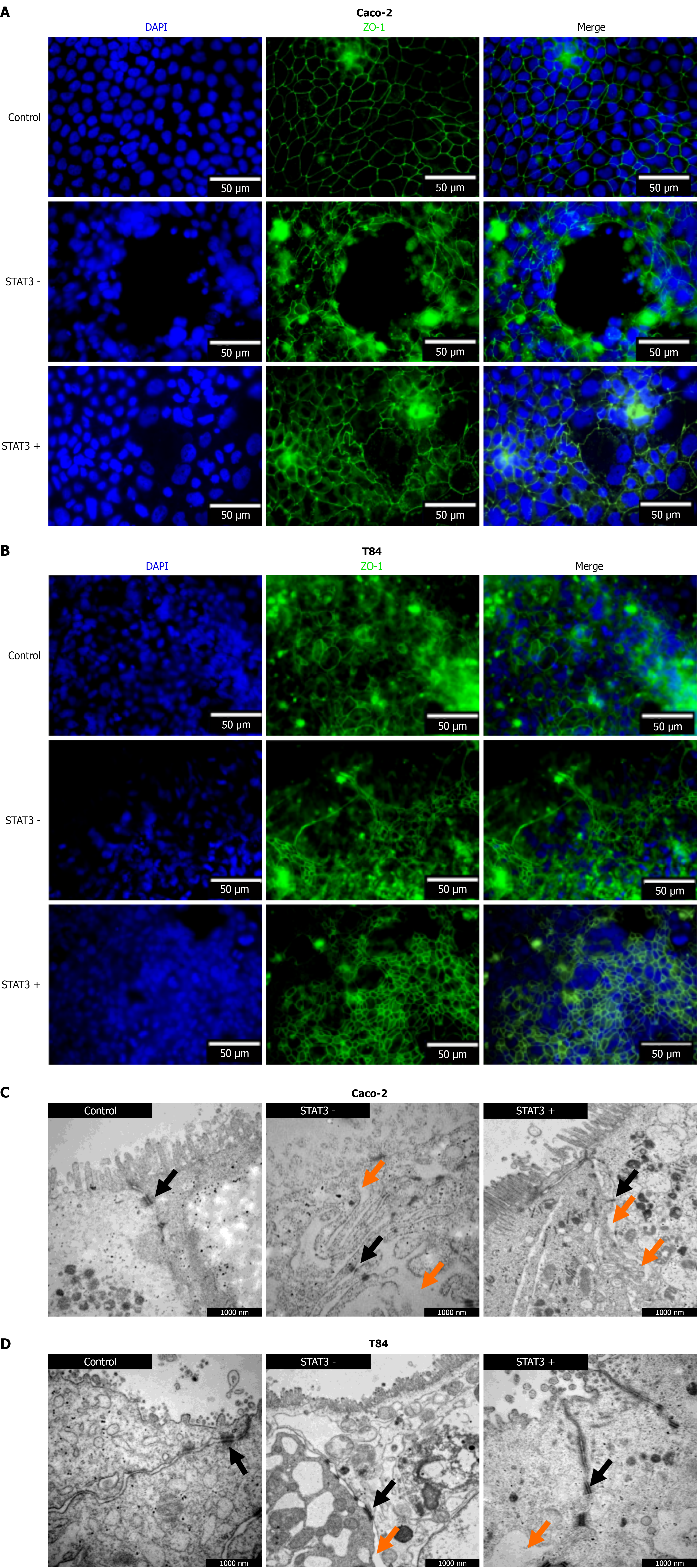
Figure 5 Immunofluorescence staining and transmission electron microscopy verify deconstructed monolayer architecture in enhanced as well as reduced signal transducer and activator of transcription 3 expressing Caco-2 and T84 monolayers.
Differential signal transducer and activator of transcription 3 (STAT3) expressing Caco-2 and T84 cell lines were grown over a period of 28 days and the formation of Caco-2 and T84 monolayers was verified during this period. A and B: Caco-2 (A) T84 (B) monolayers were fixed, permeabilized, incubated with anti zonula occludens-1, and subsequently stained with a fluorescent-labeled antibody. Nuclei were counterstained with 4’,6-diamidino-2-phenylindole. Imaging was performed using a Keyence BZ X810 fluorescence microscope; C and D: In parallel, Caco-2 (C) and T84 (D) monolayers were prepared for transmission electron microscopy (TEM). Immunofluorescence as well as TEM showed the deconstructed architecture in enhanced STAT3 (STAT3 +) as well as reduced STAT3 (STAT3 -) expressing monolayers. Orange arrows = gaps at cell boundaries, black arrows = location of tight junctions. STAT3: Signal transducer and activator of transcription 3; DAPI: 4’,6-diamidino-2-phenylindole; ZO-1: Zonula occludens-1.
- Citation: Leinz N, Beyer S, Yoganathan-Kugarajan B, Kraus N, Ortiz C, Hahnefeld L, Gurke R, Başoğlu M, Plotz G, Eimer S, Zeuzem S, Trebicka J, Welsch C, Brieger A. Dysregulated signal transducer and activator of transcription 3 drives intestinal permeability and may contribute to acute-on-chronic liver failure. World J Gastroenterol 2026; 32(30): 119465
- URL: https://www.wjgnet.com/1007-9327/full/v32/i30/119465.htm
- DOI: https://dx.doi.org/10.3748/wjg.v32.i30.119465