
















Copyright: ©Author(s) 2026.
World J Gastroenterol. Aug 14, 2026; 32(30): 119465
Published online Aug 14, 2026. doi: 10.3748/wjg.v32.i30.119465
Published online Aug 14, 2026. doi: 10.3748/wjg.v32.i30.119465
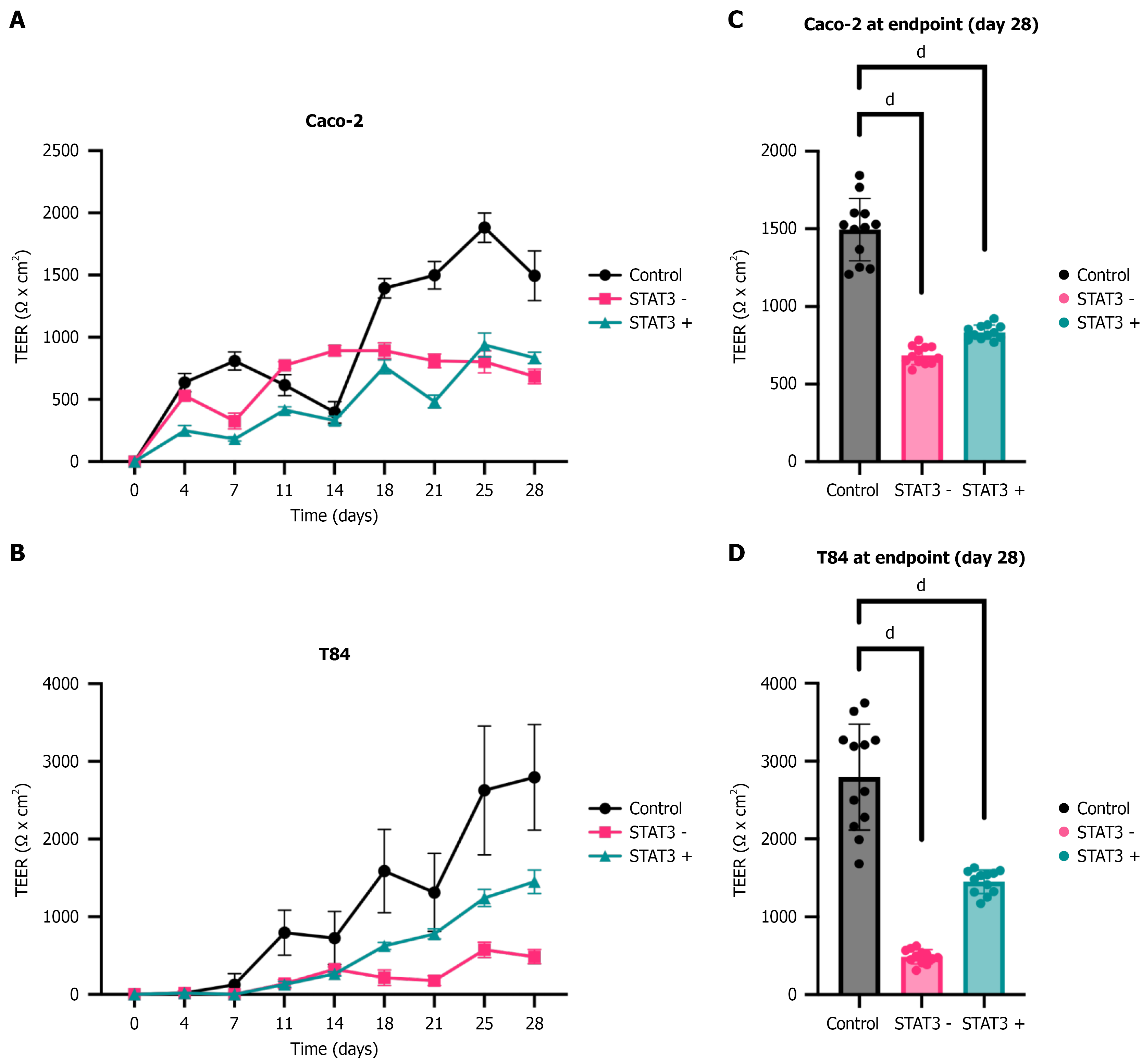
Figure 3 Signal transducer and activator of transcription 3 reduction as well as enhanced signal transducer and activator of transcription 3 expression impairs permeability of epithelial cell layers.
To determine the influence of signal transducer and activator of transcription 3 (STAT3) on epithelial permeability, monolayers consisting of Caco-2 or T84 cells with different levels of STAT3 expression were generated and epithelial permeability was analyzed by transepithelial electrical resistance (TEER) measurements over 28 days and compared to controls. A and B: Graphs show the differences of TEER values after 4 days, 7 days, 11 days, 14 days, 18 days, 21 days, 25 days and 28 days compared with endogenous STAT3 expressing (pLKO.1 transduced) controls; C and D: TEER values at day 28 were calculated in relation to controls in percent and shown as bar diagrams. Reduced (STAT3 -) as well as enhanced STAT3 (STAT3 +) expression resulted in a significant reduction in monolayer resistance values of Caco-2 and T84 monolayers. Data are expressed as mean ± SEM as appropriate. Differences between mean resistance values were assessed for statistical significance using one-way analysis of variance followed by Dunnett multiple comparisons test. P values are two-sided (n = 3-5). dP < 0.0001. STAT3: Signal transducer and activator of transcription 3; TEER: Transepithelial electrical resistance.
- Citation: Leinz N, Beyer S, Yoganathan-Kugarajan B, Kraus N, Ortiz C, Hahnefeld L, Gurke R, Başoğlu M, Plotz G, Eimer S, Zeuzem S, Trebicka J, Welsch C, Brieger A. Dysregulated signal transducer and activator of transcription 3 drives intestinal permeability and may contribute to acute-on-chronic liver failure. World J Gastroenterol 2026; 32(30): 119465
- URL: https://www.wjgnet.com/1007-9327/full/v32/i30/119465.htm
- DOI: https://dx.doi.org/10.3748/wjg.v32.i30.119465