
















Copyright: ©Author(s) 2026.
World J Gastroenterol. Aug 14, 2026; 32(30): 119177
Published online Aug 14, 2026. doi: 10.3748/wjg.119177
Published online Aug 14, 2026. doi: 10.3748/wjg.119177
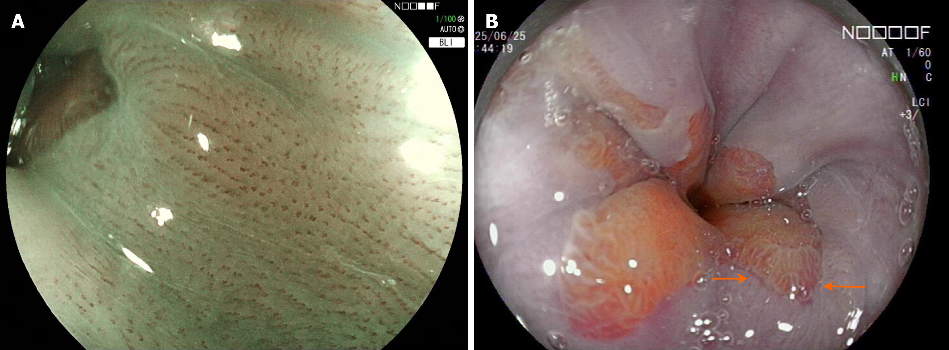
Figure 2 Subtle changes of non-erosive reflux disease under blue laser imaging and linked color imaging.
A: Increased number, dilation, and tortuosity of intrapapillary capillary loops under blue laser imaging combined with magnifying endoscopy; B: Reddish mucosal changes and prominent gastric mucosal folds observed at the squamocolumnar junction under linked color imaging. BLI: Blue laser imaging; LCI: Linked color imaging.
- Citation: Zhang WT, Xu M, Zheng Y, Wu J, Zheng YJ, Wei J, Kang T, Jiang J, Cheng Y. Endoscopic diagnosis of non-erosive reflux disease. World J Gastroenterol 2026; 32(30): 119177
- URL: https://www.wjgnet.com/1007-9327/full/v32/i30/119177.htm
- DOI: https://dx.doi.org/10.3748/wjg.119177