
















©The Author(s) 2026.
World J Gastroenterol. Jan 21, 2026; 32(3): 114226
Published online Jan 21, 2026. doi: 10.3748/wjg.v32.i3.114226
Published online Jan 21, 2026. doi: 10.3748/wjg.v32.i3.114226
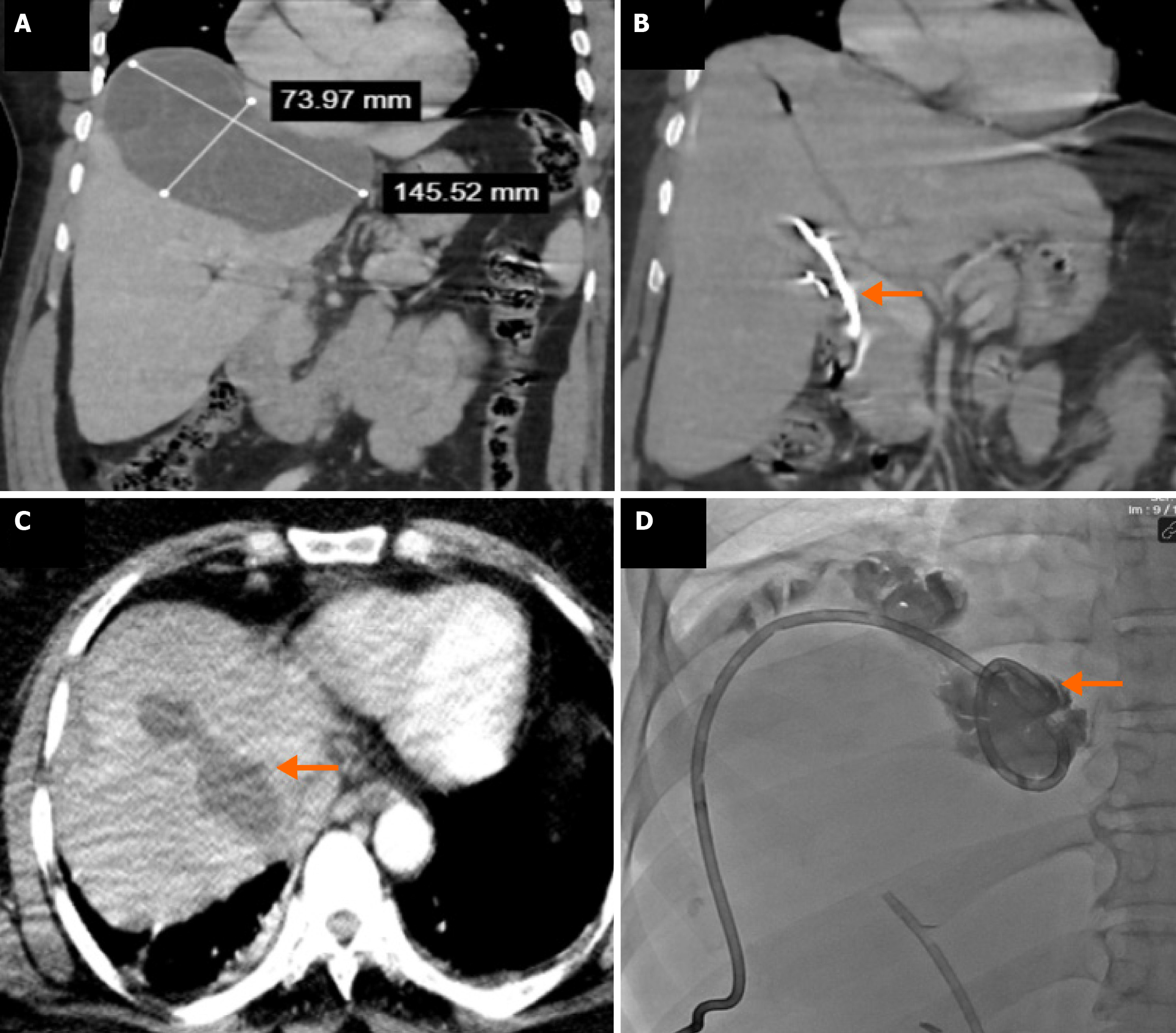
Figure 7 Pre- and postoperative computed tomography and fluoroscopy images of a 40-year-old female patient with a cystic echi
- Citation: Tahtabasi M, Kaya E, Yalcin M, Kaya V. Percutaneous vs surgical management of World Health Organization cystic echinococcosis 1 and 3a liver hydatid cysts. World J Gastroenterol 2026; 32(3): 114226
- URL: https://www.wjgnet.com/1007-9327/full/v32/i3/114226.htm
- DOI: https://dx.doi.org/10.3748/wjg.v32.i3.114226