
















©The Author(s) 2026.
World J Gastroenterol. Jan 21, 2026; 32(3): 114226
Published online Jan 21, 2026. doi: 10.3748/wjg.v32.i3.114226
Published online Jan 21, 2026. doi: 10.3748/wjg.v32.i3.114226
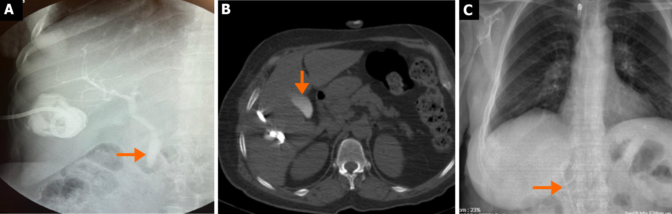
Figure 5 Images of a 54-year-old female patient with cystobiliary fistula development.
A: The cystography image of the patient with the presence of bile in drainage fluid on the second day after percutaneous treatment, shows that the contrast medium having passed into the biliary tract (arrow) and the presence of a cystobiliary fistula; B: Computed tomography image taken on the third day shows that opaque material has accumulated in the gallbladder (arrow); C: Due to increased fistula flow, papillotomy and common bile duct stenting (arrow) were performed.
- Citation: Tahtabasi M, Kaya E, Yalcin M, Kaya V. Percutaneous vs surgical management of World Health Organization cystic echinococcosis 1 and 3a liver hydatid cysts. World J Gastroenterol 2026; 32(3): 114226
- URL: https://www.wjgnet.com/1007-9327/full/v32/i3/114226.htm
- DOI: https://dx.doi.org/10.3748/wjg.v32.i3.114226