
















©The Author(s) 2026.
World J Gastroenterol. Jan 21, 2026; 32(3): 113935
Published online Jan 21, 2026. doi: 10.3748/wjg.v32.i3.113935
Published online Jan 21, 2026. doi: 10.3748/wjg.v32.i3.113935
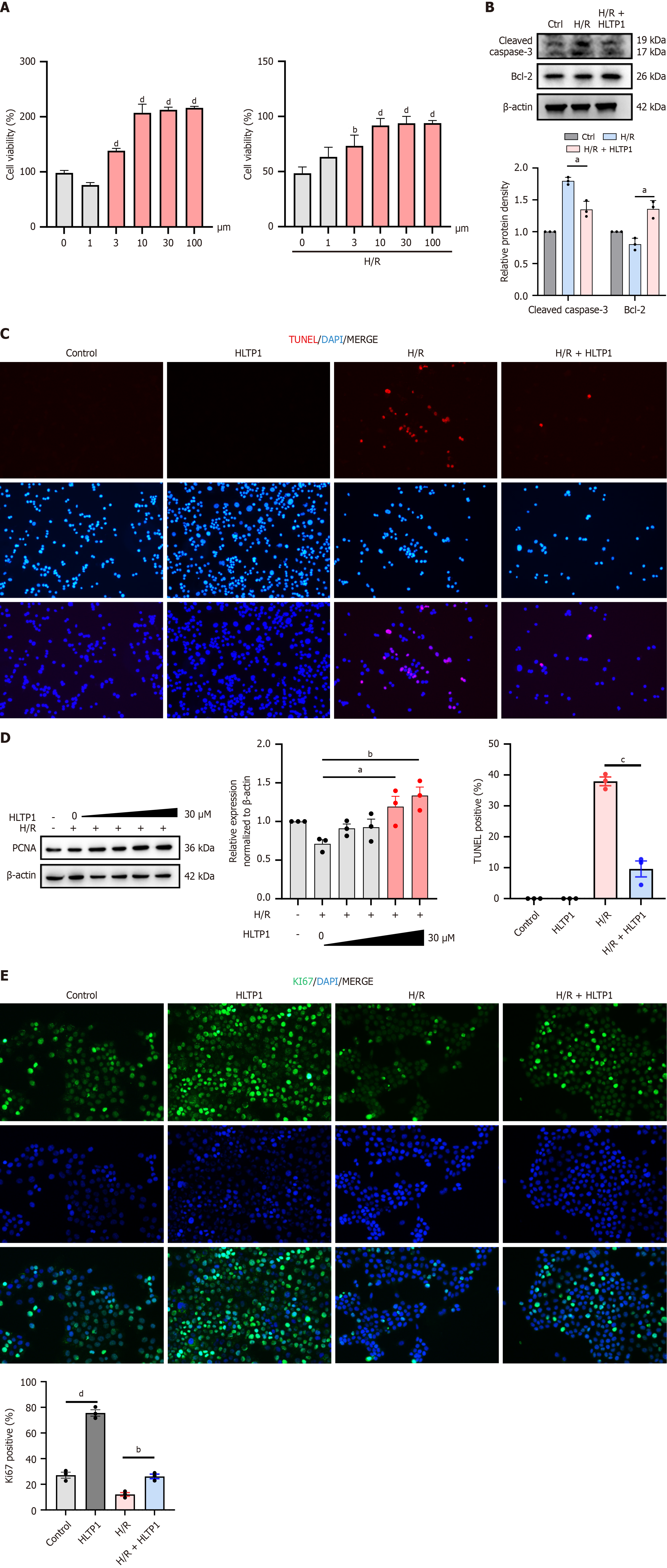
Figure 4 Human liver transplantation peptide 1 attenuates AML12 cells injury under hypoxia/reoxygenation treatment.
A: Cell viability after human liver transplantation peptide 1 treatment; B: Cleaved caspase-3 and Bcl-2 protein expression by western blotting; C: Representative images of terminal deoxynucleotidyl transferase dUTP nick end labeling staining (red) performed on AML12 cells. Nuclei are counterstained with DAPI (blue); D: PCNA protein expression by western blotting; E: Representative immunofluorescence images of AML12 cells stained for the proliferation marker Ki67 (green). Nuclei are counterstained with DAPI (blue). H/R: Hypoxia/reoxygenation; HLTP1: Human liver transplantation peptide 1; TUNEL: Terminal deoxynucleotidyl transferase dUTP nick end labeling. aP < 0.05, bP < 0.01, cP < 0.001, and dP < 0.0001.
- Citation: Xie HW, Bao Q, Chen ZX, Zhang XM, Liu XY, Wang R, Cai YS, Sun P. Targeting Jun N-terminal kinase phosphorylation: A human-derived hepatoprotective peptide human liver transplantation peptide 1 attenuates hepatic ischemia-reperfusion injury. World J Gastroenterol 2026; 32(3): 113935
- URL: https://www.wjgnet.com/1007-9327/full/v32/i3/113935.htm
- DOI: https://dx.doi.org/10.3748/wjg.v32.i3.113935