
















Copyright: ©Author(s) 2026.
World J Gastroenterol. Aug 7, 2026; 32(29): 120498
Published online Aug 7, 2026. doi: 10.3748/wjg.120498
Published online Aug 7, 2026. doi: 10.3748/wjg.120498
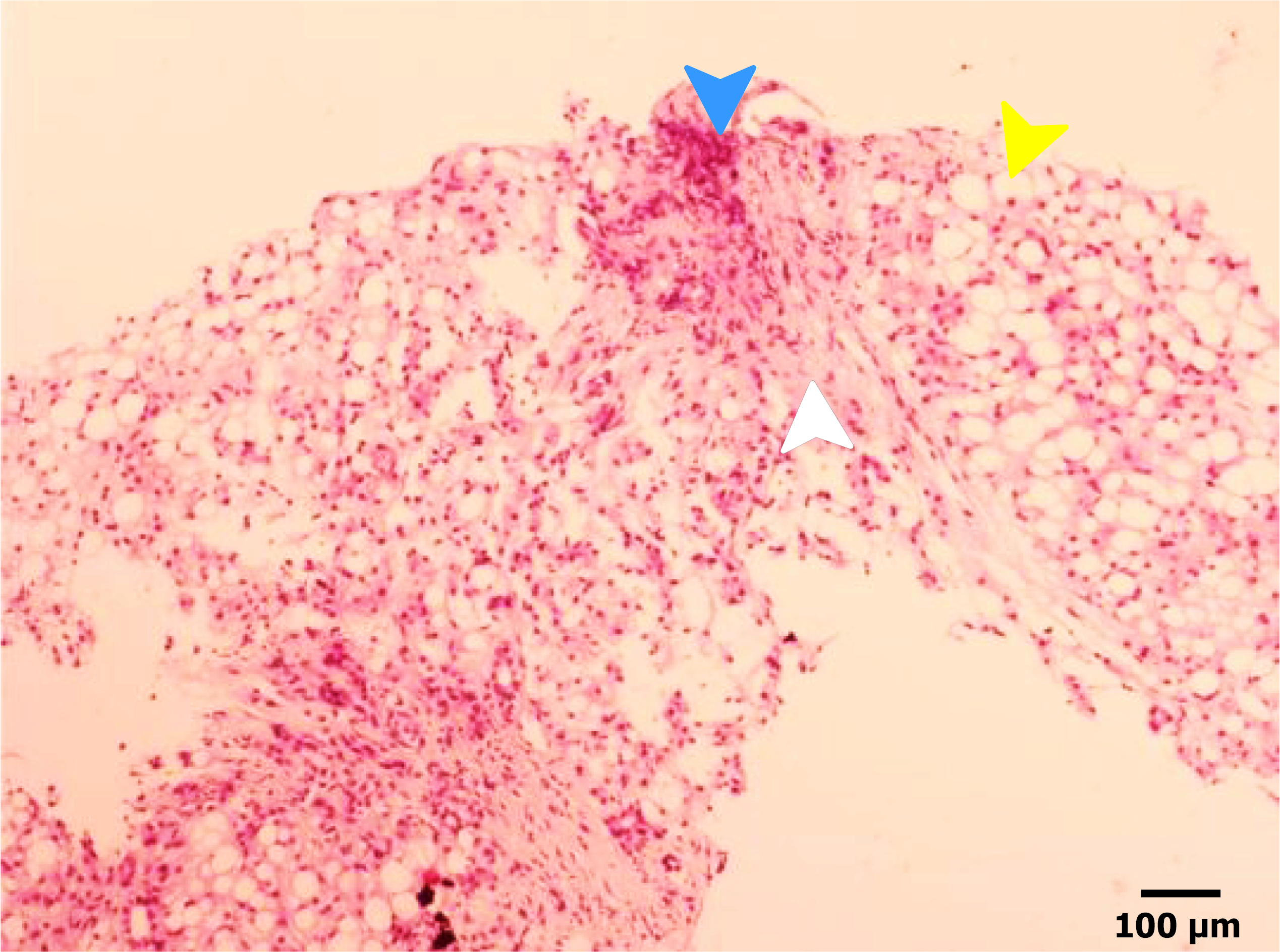
Figure 3 Histopathological examination of the liver biopsy specimen obtained at the 4-year postoperative follow-up (hematoxylin and eosin staining).
The liver tissue shows diffuse macrovesicular steatosis of hepatocytes (yellow arrow), accompanied by inflammatory cell infiltration predominantly composed of lymphocytes (blue arrow). Marked fibrous tissue proliferation is observed within the lobules and portal areas (white arrow), resulting in blurring of the normal lobular architecture. In addition, increased proliferation of small bile ducts is noted within hepatocytes and portal tracts, indicating advanced chronic liver injury with fibrotic remodeling.
- Citation: Hu H, Liu YJ, Li MC, Li Y, Lin SD. Rapid progression to liver failure following pancreatoduodenectomy in an adolescent: A case report. World J Gastroenterol 2026; 32(29): 120498
- URL: https://www.wjgnet.com/1007-9327/full/v32/i29/120498.htm
- DOI: https://dx.doi.org/10.3748/wjg.120498