
















Copyright: ©Author(s) 2026.
World J Gastroenterol. Aug 7, 2026; 32(29): 119920
Published online Aug 7, 2026. doi: 10.3748/wjg.119920
Published online Aug 7, 2026. doi: 10.3748/wjg.119920
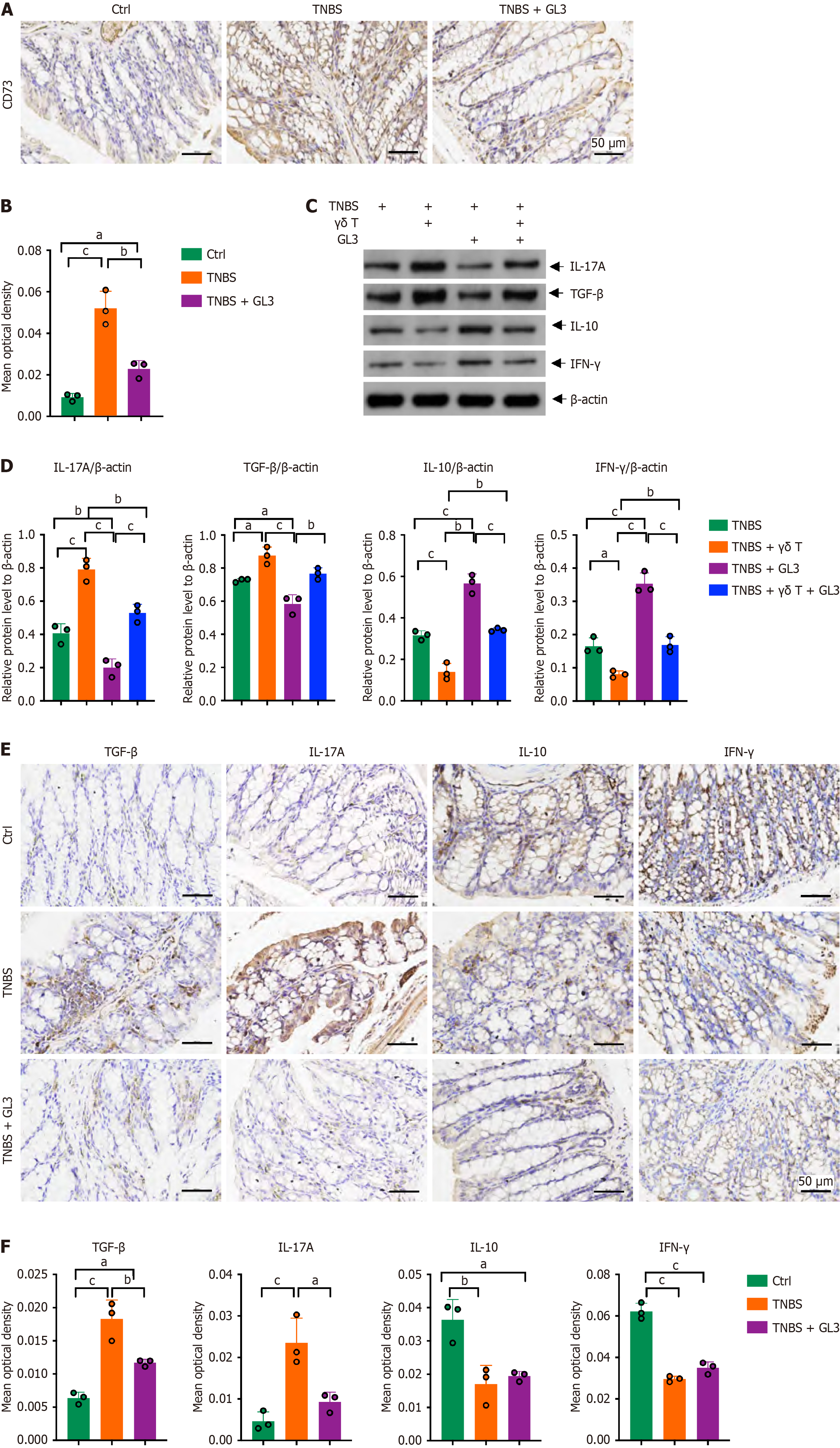
Figure 3 CD73 was downregulated after γδ T cells depletion in mice with intestinal fibrosis.
A and B: Immunohistochemistry (IHC) staining of CD73 and the quantification of average optical density (OD) of CD73 in the colon tissues. Quantification of IHC staining expressed as mean OD. Scale bars: 50 μm; C and D: Western blot and the quantification result of the expression levels of interleukin (IL)-17A, transforming growth factor-beta, interferon γ and IL10 using colon tissues in each group (n = 3); E and F: IHC staining of IL-17A, transforming growth factor-beta, interferon γ and IL10 and the quantification of average OD of these proteins in the colon tissues. Quantification of IHC staining expressed as mean OD. Scale bars: 50 μm. Data were presented as mean ± SD. aP < 0.05, bP < 0.01, cP < 0.001. TNBS: 2,4,6-trinitrobenzenesulfonic acid; IL: Interleukin; TGF: Transforming growth factor; IFN: Interferon.
- Citation: Dong LW, Ma ZC, Fu J, Huang BL, Liu FJ, Liang DC, Sun DM, Lan C. γδ T cells exacerbate intestinal fibrosis in mice with experimental colitis via activation of the CD73-adenosine receptor-cyclic AMP signaling pathway. World J Gastroenterol 2026; 32(29): 119920
- URL: https://www.wjgnet.com/1007-9327/full/v32/i29/119920.htm
- DOI: https://dx.doi.org/10.3748/wjg.119920