
















Copyright: ©Author(s) 2026.
World J Gastroenterol. Aug 7, 2026; 32(29): 119374
Published online Aug 7, 2026. doi: 10.3748/wjg.119374
Published online Aug 7, 2026. doi: 10.3748/wjg.119374
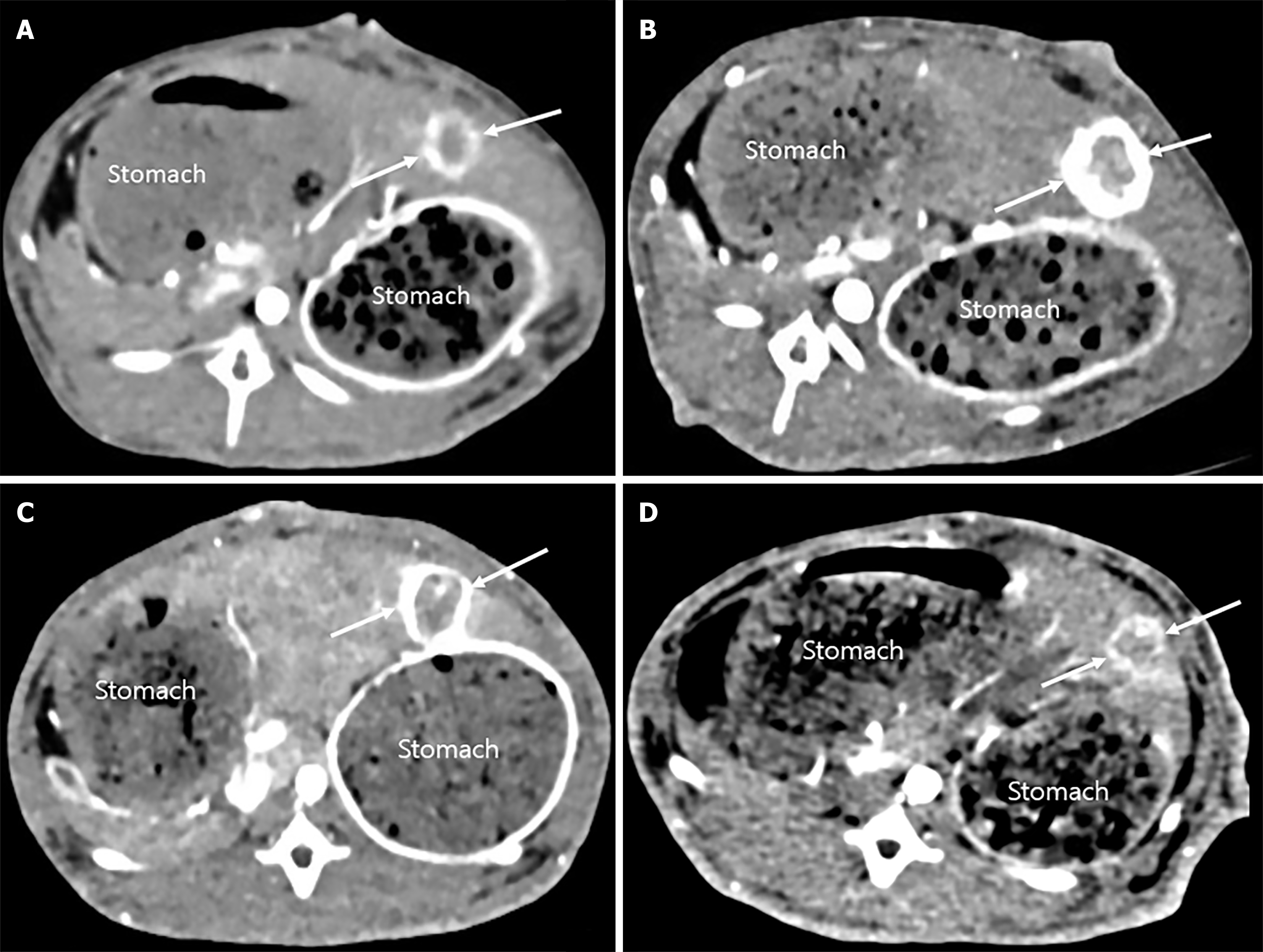
Figure 2 Representative computed tomography images of VX2 liver tumors before and after transcatheter arterial chemoembolization.
A and B: In the normal saline with doxorubicin group, pre-transcatheter arterial chemoembolization (TACE) (A) and post-TACE (B) images show a marked enlargement in tumor volume (arrows) and a significantly higher tumor growth ratio; C and D: In the triolein emulsion with doxorubicin group, pre-TACE (C) and post-TACE (D) images demonstrate a smaller reduction in tumor volume (arrows) and a significantly lower tumor growth ratio. All panels depict the largest tumor diameter slice for accurate volume estimation, and the tumor locations are consistent across pre- and post-TACE images.
- Citation: Kim YW, Kim HJ, Choi SH, Yun MS. Enhancing delivery of doxorubicin using triolein emulsion in rabbit VX2 liver tumor model. World J Gastroenterol 2026; 32(29): 119374
- URL: https://www.wjgnet.com/1007-9327/full/v32/i29/119374.htm
- DOI: https://dx.doi.org/10.3748/wjg.119374