
















Copyright: ©Author(s) 2026.
World J Gastroenterol. Aug 7, 2026; 32(29): 119374
Published online Aug 7, 2026. doi: 10.3748/wjg.119374
Published online Aug 7, 2026. doi: 10.3748/wjg.119374
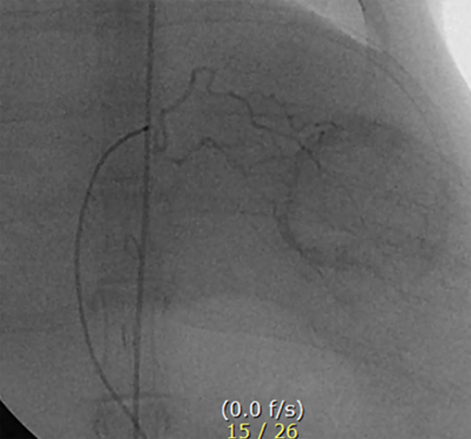
Figure 1 Angiography of VX2 tumor via the hepatic artery in a rabbit liver.
The hepatic angiogram demonstrates tumor staining in the left lobe of the liver. The catheter tip is positioned in the left hepatic artery through the celiac axis using the trans-auricular approach. Triolein emulsion or normal saline, along with doxorubicin, were administered through a microcatheter.
- Citation: Kim YW, Kim HJ, Choi SH, Yun MS. Enhancing delivery of doxorubicin using triolein emulsion in rabbit VX2 liver tumor model. World J Gastroenterol 2026; 32(29): 119374
- URL: https://www.wjgnet.com/1007-9327/full/v32/i29/119374.htm
- DOI: https://dx.doi.org/10.3748/wjg.119374