
















Copyright: ©Author(s) 2026.
World J Gastroenterol. Jul 28, 2026; 32(28): 118939
Published online Jul 28, 2026. doi: 10.3748/wjg.118939
Published online Jul 28, 2026. doi: 10.3748/wjg.118939
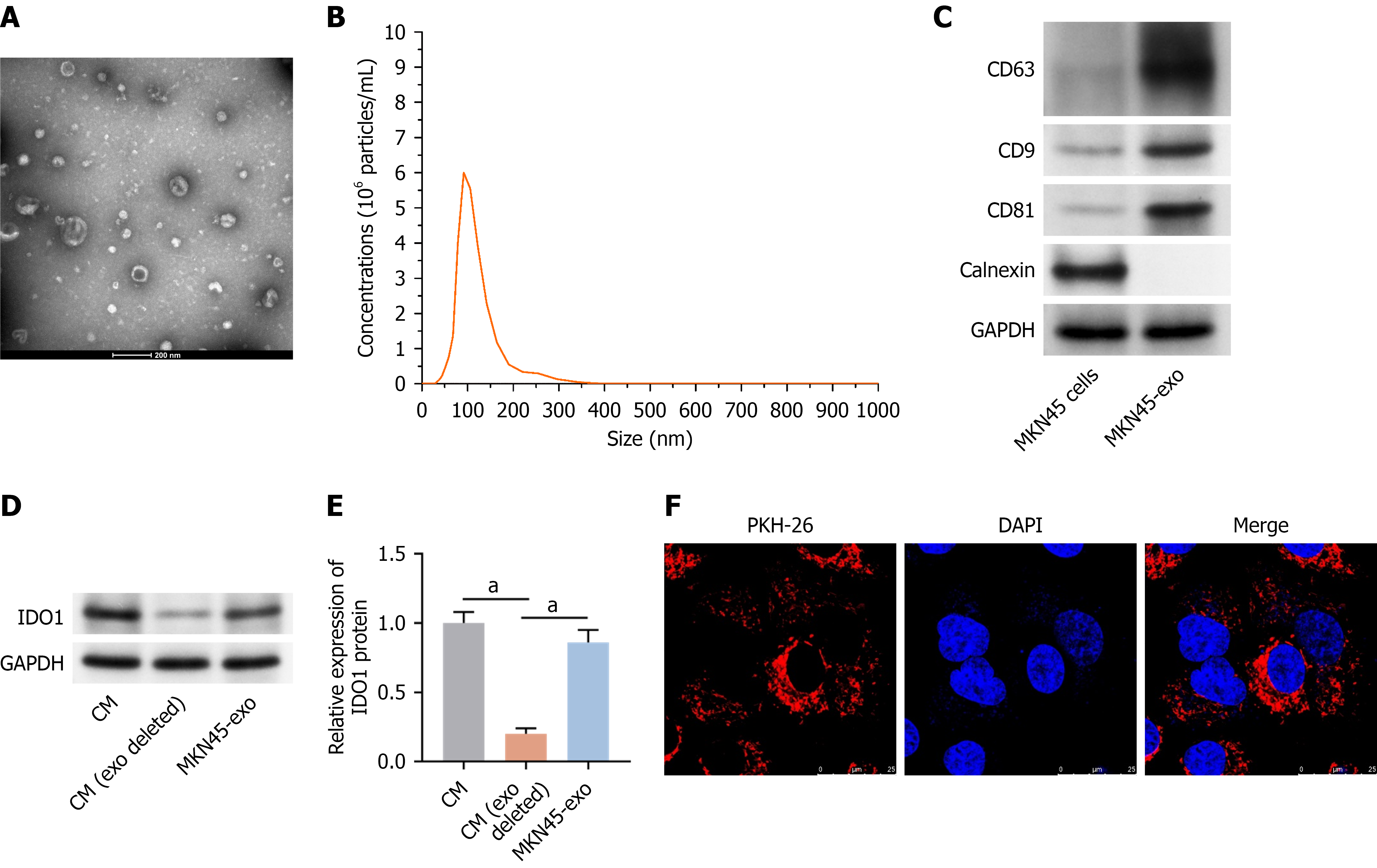
Figure 2 Indoleamine 2,3 dioxygenase 1 was highly enriched in exosomes derived from gastric cancer cells.
Exosomes were isolated from MKN45 cells. A: Exosome morphology was examined using transmission electron microscopy; B: Nanoparticle tracking analysis was employed to determine size distribution; C: The presence of exosomal markers was confirmed using Western blot analysis; D and E: Indoleamine 2,3 dioxygenase 1 expression in exosomes was measured using Western blot analysis; F: Uptake of PKH26-labeled exosomes by dendritic cells was visualized using microscopy. Data were presented as mean ± SD. aP < 0.01. CD: Cluster of differentiation; CM: Conditioned medium; DAPI: 4’,6-diamidino-2-phenylindole; exo: Exosomal; GAPDH: Glyceraldehyde-3-phosphate dehydrogenase; IDO1: Indoleamine 2,3 dioxygenase 1.
- Citation: Liu YY, Ai JH, Zeng LM, Liu XN, Wu F. Gastric cancer-derived exosomal indoleamine 2,3 dioxygenase 1 drives immunosuppression in gastric cancer by suppressing dendritic cell maturation. World J Gastroenterol 2026; 32(28): 118939
- URL: https://www.wjgnet.com/1007-9327/full/v32/i28/118939.htm
- DOI: https://dx.doi.org/10.3748/wjg.118939