
















Copyright: ©Author(s) 2026.
World J Gastroenterol. Jul 28, 2026; 32(28): 118458
Published online Jul 28, 2026. doi: 10.3748/wjg.118458
Published online Jul 28, 2026. doi: 10.3748/wjg.118458
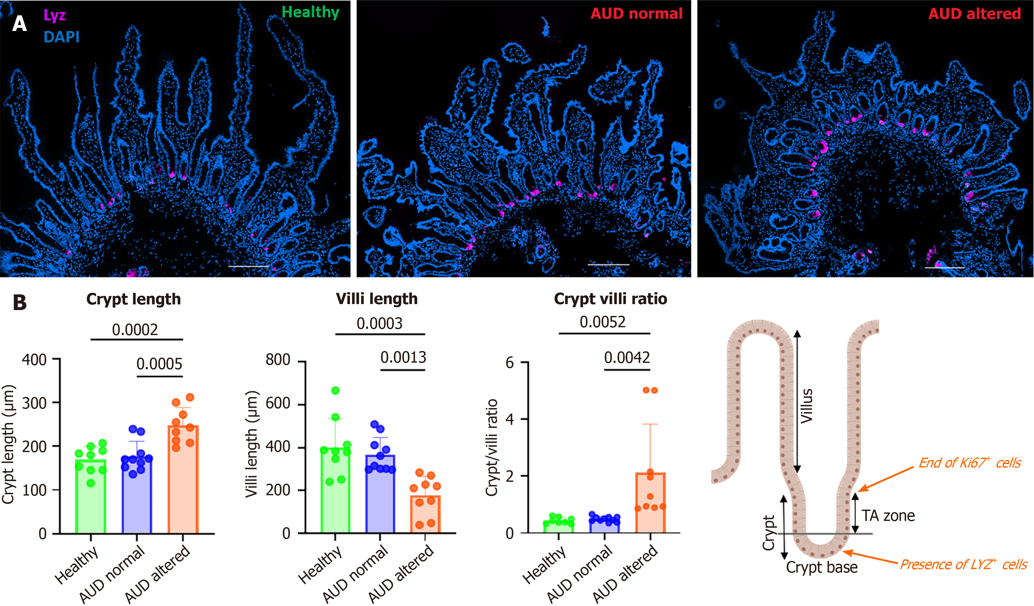
Figure 1 Crypt/villus morphology in alcohol use disorder patients.
A: Representative immunofluorescence staining of duodenal biopsies showing morphology alterations in a subset of alcohol use disorder (AUD) patients (AUD altered) compared to healthy subjects and AUD patients with normal morphology (AUD normal). Lysozyme in purple stains Paneth cells to identify the crypt base. DAPI (blue) is used as counterstaining to show the nucleus; B: Quantification of the crypt (μm) and villi length (μm). Five to ten crypts/villi were measured per patient. Data are presented as mean ± SD. Crypt and villus length are significantly modified in the AUD altered group resulting in a higher crypt to villi ratio in these patients. The schematic illustration shows the boundaries of the crypt, the transit-amplifying zone, and the villus that defined the limits for the measurements. Healthy (n = 9); AUD normal (n = 10); AUD altered (n = 9). AUD: Alcohol use disorder.
- Citation: Toulehohoun AG, Ambroise J, Illiyas B, Maccioni L, Lupianez J, Manco R, Clerbaux LA, Bouzin C, Schnabl B, Stärkel P. Impairment of the intestinal epithelium barrier through alteration of proliferation and differentiation balance in alcohol use disorder. World J Gastroenterol 2026; 32(28): 118458
- URL: https://www.wjgnet.com/1007-9327/full/v32/i28/118458.htm
- DOI: https://dx.doi.org/10.3748/wjg.118458