
















Copyright: ©Author(s) 2026.
World J Gastroenterol. Jul 21, 2026; 32(27): 118794
Published online Jul 21, 2026. doi: 10.3748/wjg.118794
Published online Jul 21, 2026. doi: 10.3748/wjg.118794
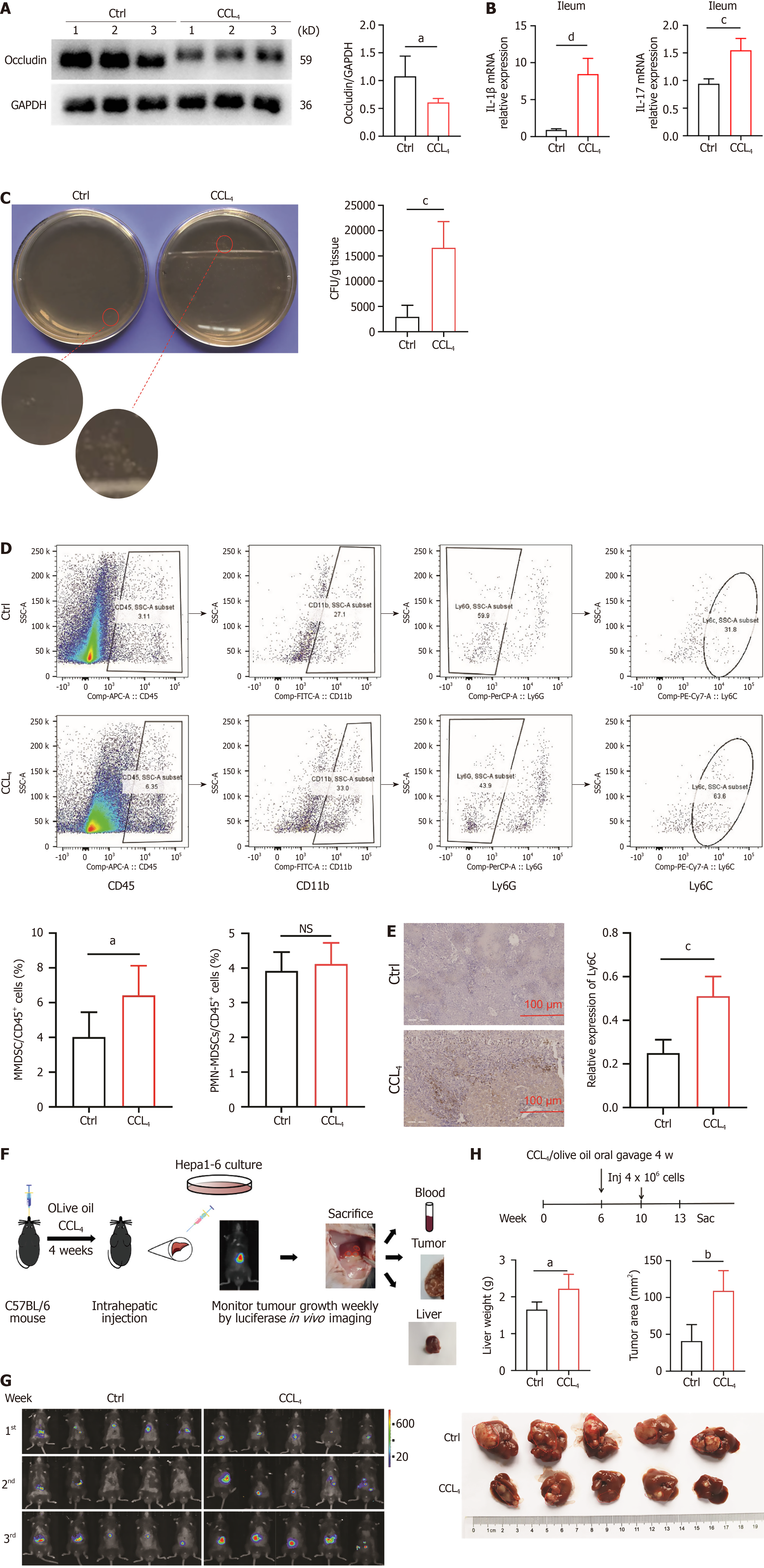
Figure 4 An impaired intestinal mucosal barrier renders the liver susceptible to exposure to gut microbiota and promotes hepatocellular carcinoma growth.
A: Occludin relative expression in ileum in control (Ctrl) and CCL4 groups; B: Interleukin-1β and interleukin-17 mRNA expression in ileum in Ctrl and CCL4 groups (n = 5 for each group); C: Hepatocellular carcinoma (HCC) tissues from Ctr and CCL4 mice were homogenized and cultured on a BHI medium without antibiotics. The clone formation units were calculated; D: The flow cytometry-based proportional changes of monocytic myeloid-derived suppressor cells and polymorphonuclear-myeloid-derived suppressor cells in the tumor microenvironment (TME) between Ctrl and CCl4 groups; E: The Ly6C relative expression in TME in Ctrl and CCL4 groups; F: Establishment of murine HCC models and monitoring tumor growth; G: Weekly in vivo imaging was performed to monitor tumor growth in Ctrl and CCL4 groups; H: Following the schematic diagram of animal interventions, tumor size and liver weight changes were compared between the Ctrl and CCl4 groups post-intervention. aP < 0.05, bP < 0.01; bP < 0.001; cP < 0.0001; NS: Not significant. IL: Interleukin; CFU: Clone formation unit; PMN: Polymorphonuclear; MDSC: Myeloid-derived suppressor cell; MMDSC: Monocytic myeloid-derived suppressor cell; Ctrl: Control.
- Citation: Zhang Y, Zhou YL, Nie X, Zhang LJ, Xu WW, Wang JQ, Zhao HL, Xu JK, Xu J, Xu HM, Li JH, Huang WQ, Yang J, Yang YX, Zhan Q, Lin Y, Nie YQ. Gram-negative bacteria and endotoxins promote hepatocellular carcinoma progression through the recruitment of myeloid-derived suppressor cells. World J Gastroenterol 2026; 32(27): 118794
- URL: https://www.wjgnet.com/1007-9327/full/v32/i27/118794.htm
- DOI: https://dx.doi.org/10.3748/wjg.118794