
















Copyright: ©Author(s) 2026.
World J Gastroenterol. Jul 21, 2026; 32(27): 118794
Published online Jul 21, 2026. doi: 10.3748/wjg.118794
Published online Jul 21, 2026. doi: 10.3748/wjg.118794
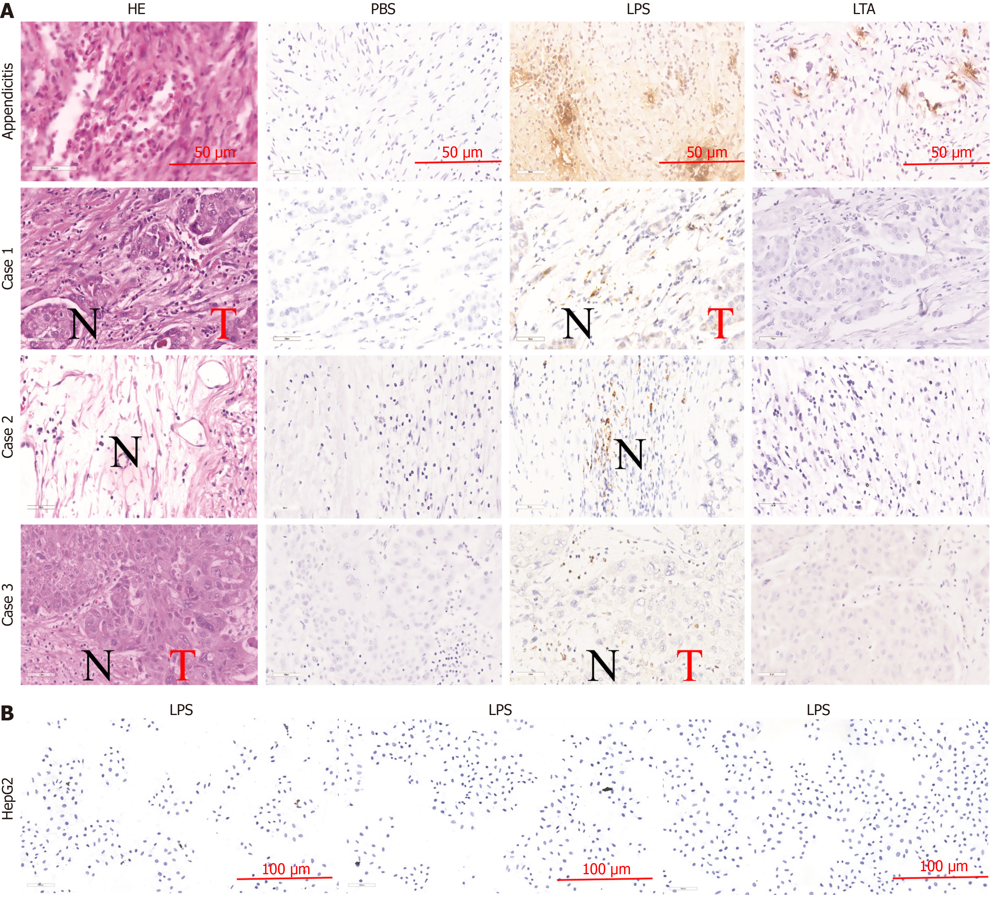
Figure 1 Lipopolysaccharide was found present in both tumor and non-tumor tissues in paraffin sections of hepatocellular carcinoma.
A: Hematoxylin-eosin staining and immunohistochemistry (50 μm) revealed the presence of lipopolysaccharide (LPS) in both tumor tissue regions and non-tumor tissue regions; B: LPS immunohistochemistry in different batches of HepG2 cells (100 μm). T: Tumor tissue region; N: Non-tumor tissue region; HE: Hematoxylin-eosin; PBS: Phosphate-buffered saline; LPS: Lipopolysaccharide; LTA: Lipoteichoic acid.
- Citation: Zhang Y, Zhou YL, Nie X, Zhang LJ, Xu WW, Wang JQ, Zhao HL, Xu JK, Xu J, Xu HM, Li JH, Huang WQ, Yang J, Yang YX, Zhan Q, Lin Y, Nie YQ. Gram-negative bacteria and endotoxins promote hepatocellular carcinoma progression through the recruitment of myeloid-derived suppressor cells. World J Gastroenterol 2026; 32(27): 118794
- URL: https://www.wjgnet.com/1007-9327/full/v32/i27/118794.htm
- DOI: https://dx.doi.org/10.3748/wjg.118794