
















Copyright: ©Author(s) 2026.
World J Gastroenterol. Jul 21, 2026; 32(27): 117897
Published online Jul 21, 2026. doi: 10.3748/wjg.117897
Published online Jul 21, 2026. doi: 10.3748/wjg.117897
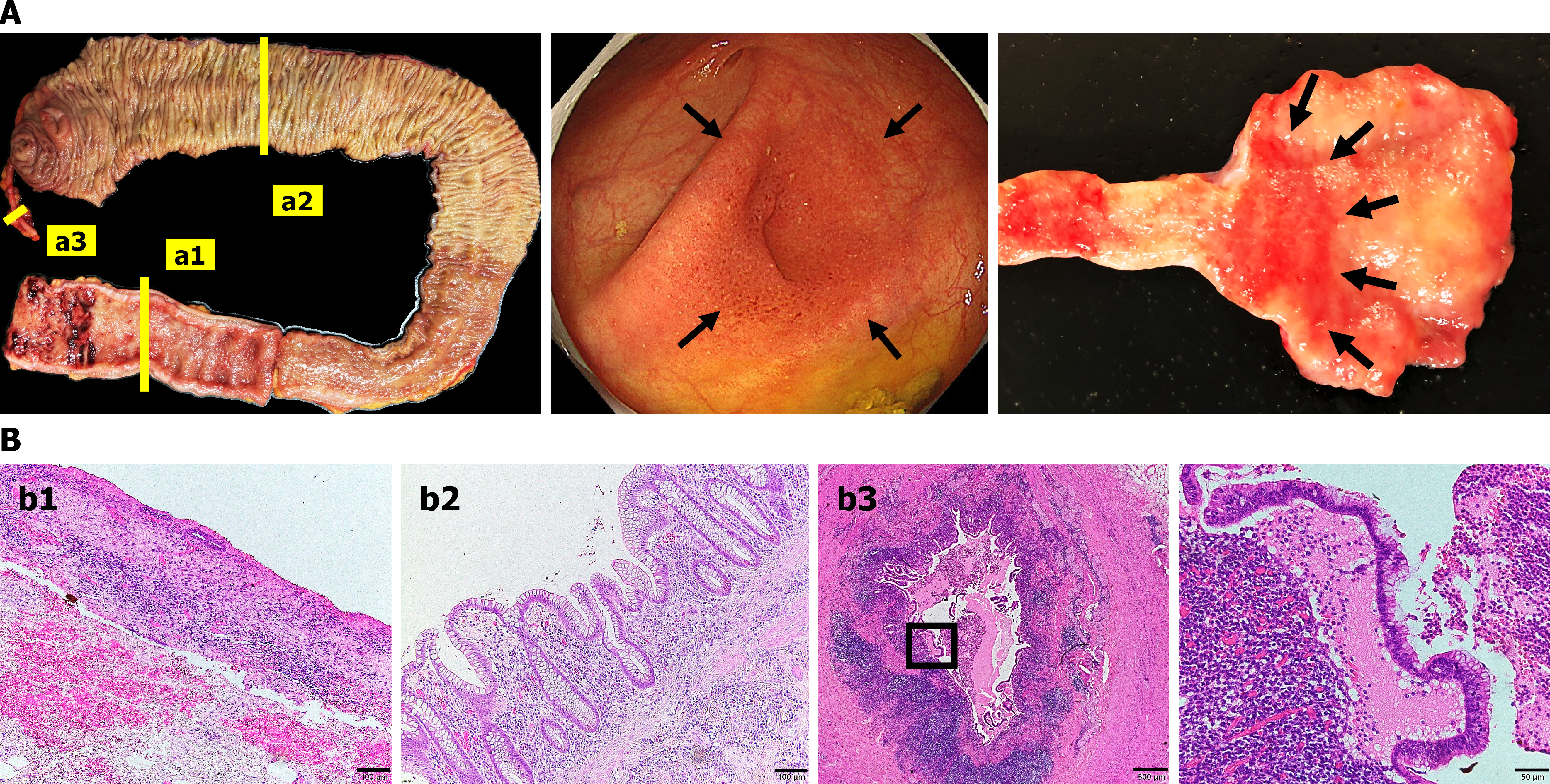
Figure 1 Appendiceal inflammation as skip lesions in ulcerative colitis.
A: The left part shows a macroscopic image of a surgical specimen from a patient with active ulcerative colitis who underwent total proctocolectomy (a1: Rectum, a2: Ascending colon, and a3: Appendix). The middle part presents endoscopic findings of the appendiceal orifice in ulcerative colitis. The right part displays a macroscopic image of the appendiceal orifice. The black arrows indicate appendiceal orifice inflammation. The stained sections in a1, a2, and a3 correspond to b1, b2, and b3, respectively; B: Hematoxylin and eosin staining of the rectum (b1), bar = 100 μm; ascending colon (b2), bar = 100 μm; appendix (b3), bars = 500 μm (left), and 50 μm [right (enlarged view of the black square in the left panel)].
- Citation: Izutani Y, Ogino T, Sakakibara S, Kishi Y, Liu YC, Nakano Y, Fukada A, Sekido Y, Kuwahara R, Kihara T, Takeda M, Hata T, Hamabe A, Miyoshi N, Uemura M, Ikeuchi H, Hirota S, Takeda K, Mizushima T, Okuzaki D, Doki Y, Eguchi H. Contribution of the appendix to ulcerative colitis by supplying pathogenic immunoglobulin G-producing cells. World J Gastroenterol 2026; 32(27): 117897
- URL: https://www.wjgnet.com/1007-9327/full/v32/i27/117897.htm
- DOI: https://dx.doi.org/10.3748/wjg.117897