
















Copyright: ©Author(s) 2026.
World J Gastroenterol. Jul 14, 2026; 32(26): 118923
Published online Jul 14, 2026. doi: 10.3748/wjg.v32.i26.118923
Published online Jul 14, 2026. doi: 10.3748/wjg.v32.i26.118923
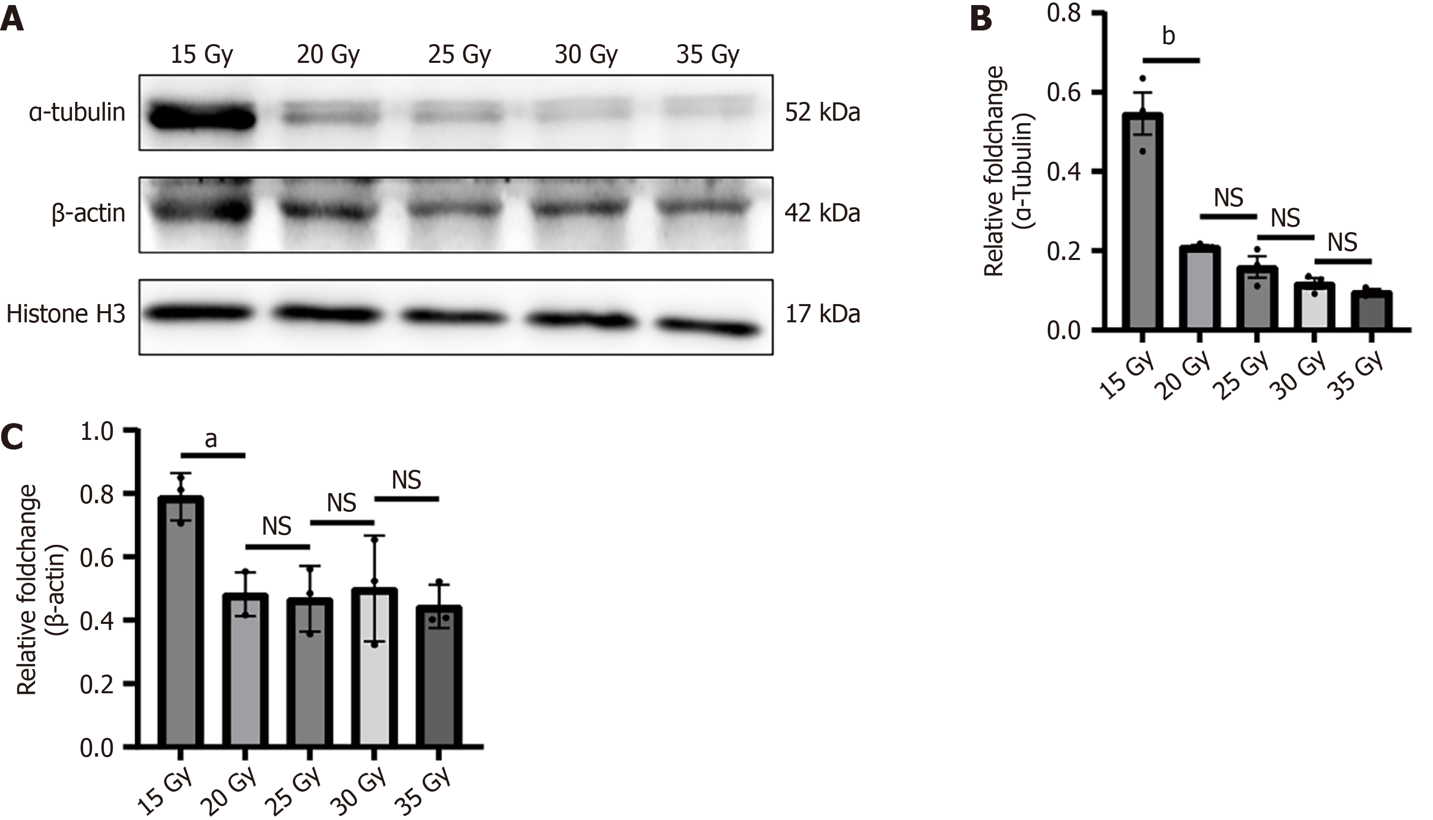
Figure 5 High-dose irradiation induces significant degradation of cytoskeletal proteins in rat rectal tissues.
A: Representative Western blot images of the cytoskeletal components α-tubulin and β-actin in rectal tissues at 2 weeks post-irradiation across different dose groups (15-35 Gy). Histone H3 was utilized as an internal loading control to represent total nuclear protein and confirm equal cell equivalent loading per lane, demonstrating stable expression across all doses; B and C: Quantitative densitometric analysis of relative α-tubulin (B) and β-actin (C) protein levels, normalized to histone H3. The data indicate a severe, dose-dependent loss of cytoskeletal integrity starting at 20 Gy. Data are presented as mean ± SD. aP < 0.05 compared to the 15 Gy group; bP < 0.0001 compared to the 15 Gy group. NS: Not significant.
- Citation: Chang Y, Sun R, Juan W, Xu Z, Xu HY, Zen K, Xuan J. Establishment of a standardized rat model of chronic radiation proctitis and preliminary evaluation of its association with tubulin. World J Gastroenterol 2026; 32(26): 118923
- URL: https://www.wjgnet.com/1007-9327/full/v32/i26/118923.htm
- DOI: https://dx.doi.org/10.3748/wjg.v32.i26.118923