
















Copyright: ©Author(s) 2026.
World J Gastroenterol. Jul 14, 2026; 32(26): 114936
Published online Jul 14, 2026. doi: 10.3748/wjg.114936
Published online Jul 14, 2026. doi: 10.3748/wjg.114936
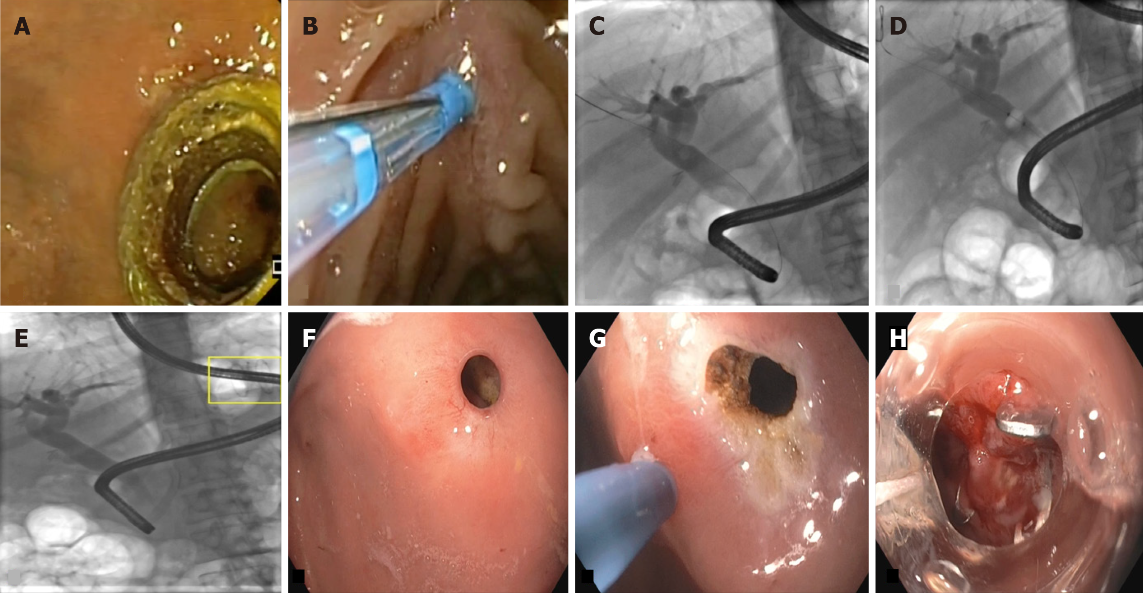
Figure 6 Endoscopic ultrasound-directed trans-gastric endoscopic retrograde cholangiopancreatography for management of biliary obstruction in altered anatomy.
A: Endoscopic image shows the presence of lumen apposing metal stent (LAMS) in-situ in the remnant gastric pouch; this is the route which is used through which the duodenoscope is passed to each of the papilla; B: Once papilla is reached, selective cannulation of bile duct is performed using a standard sphincterotome; C: Fluoroscopic image shows the guidewire in the common bile duct (CBD), following which contrast was given and cholangiogram was taken. Cholangiogram shows presence of dilated intrahepatic biliary radicles with a filling defect in CBD (likely stone); also noted are presence of metallic clips near cystic duct (indication of past laparoscopic cholecystectomy); D: Balloon sweeps were taken to clear the CBD of the stones or sludge; E: Check cholangiogram shows no filling defect in CBD; yellow box denoted the presence of LAMS through which the duodenoscope has passed; after completion of endoscopic retrograde cholangiopancreatography, LAMS was removed in same session; F: Follow up endoscopy after a month revealed persistent gastro-gastrostomy fistula; G: Margins were made raw by argon plasma coagulation; H: The fistula was closed using an 11/6t over-the-scope clip.
- Citation: Mishra S, Dhar J, Crinò SF, Samanta J. Technical overview of difficult biliary cannulation in endoscopic retrograde cholangiopancreatography: A guide for the endoscopist. World J Gastroenterol 2026; 32(26): 114936
- URL: https://www.wjgnet.com/1007-9327/full/v32/i26/114936.htm
- DOI: https://dx.doi.org/10.3748/wjg.114936